
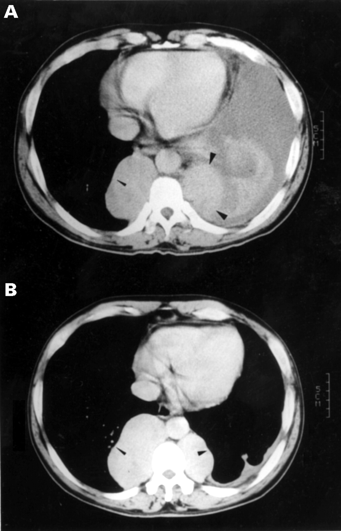
Figure 2
Computed tomographic scan of the chest showing (A) massive left sided pleural effusion and multiple lobulated paravertebral masses with well enhanced contrast (arrowheads) and (B) one year later only multiple lobulated paravertebral masses (arrowhead) were found.
Vol 79 Issue 5
Table of Contents


Share this article
Click the icon of the social media platform on which you would like to share this article.


Email this article to a friend
Respond to this article