Article Text

Statistics from Altmetric.com
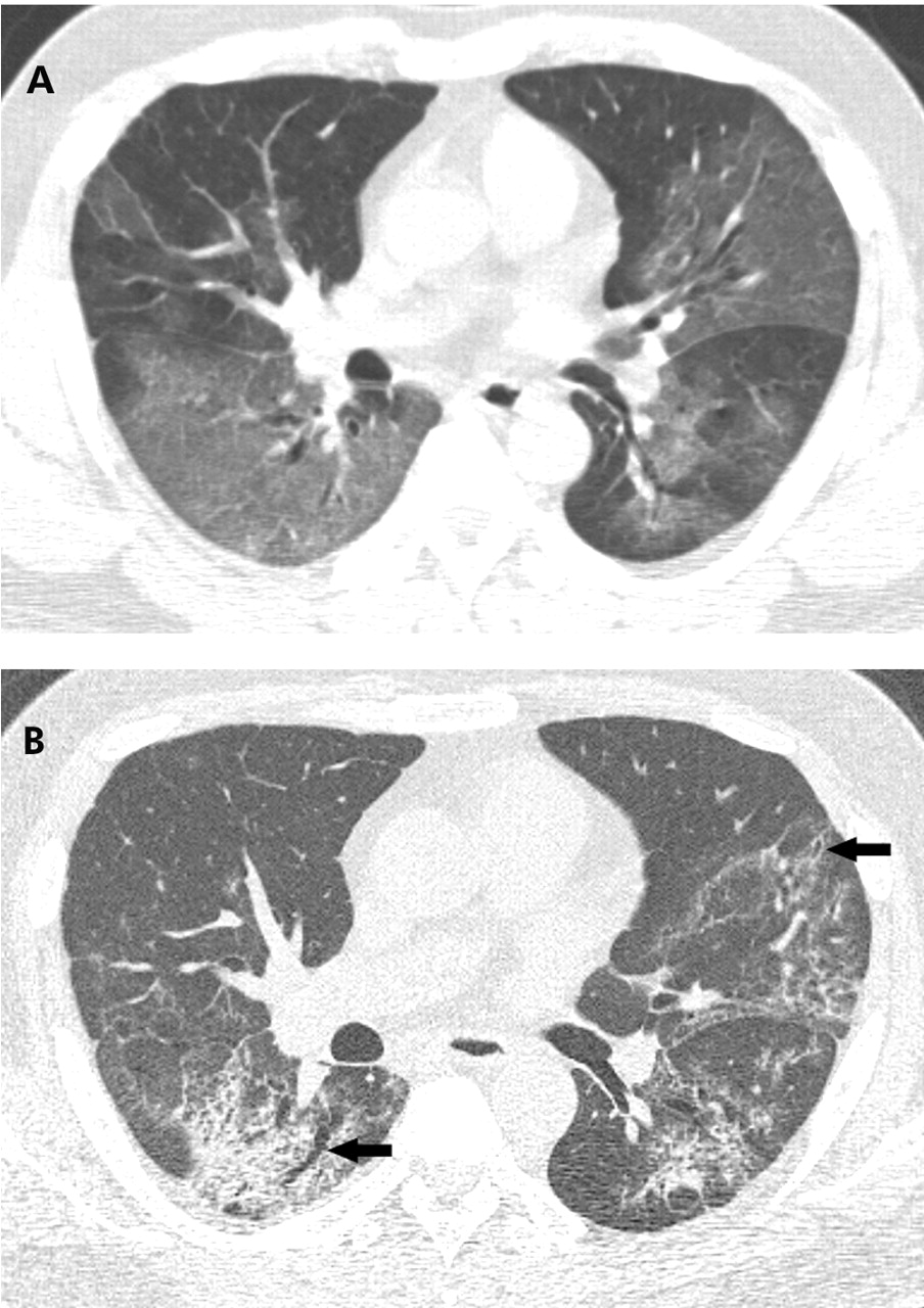
The radiological manifestations of severe acute respiratory syndrome (SARS) typically consist of focal unilateral or bilateral areas of hazy increased density (ground glass opacities) or airspace consolidation (fig 1A).1 In most patients the abnormalities gradually improve over several days following treatment. Approximately 20–25% of patients, however, show progressive deterioration with the development of confluent bilateral areas of consolidation.2 These patients frequently develop acute respiratory distress syndrome or have a protracted clinical course. Necroscopic examination of patients with SARS has shown features of diffuse alveolar damage. Patients with residual clinical symptoms 2 or more weeks after initial presentation often have a reticular pattern visible on the radiograph and irregular lines, architectural distortion, and traction bronchiectasis evident on the high resolution CT scan (fig 1B). These findings suggest the presence of fibrosis. Long term follow up will be required to determine the prevalence of fibrosis in patients who recover from SARS.

A 48 year old man with SARS. (A) High resolution CT scan performed 12 hours after hospital admission showing extensive bilateral ground glass opacities. (B) High resolution CT scan performed 27 days after admission showing fine reticular pattern superimposed on ground glass pattern. Note also distortion of the lung architecture and dilatation and irregular contour of the bronchi (traction bronchiectasis; arrows) in areas with reticulation. These findings suggest the presence of fibrosis.
Learning points
-
Characteristic radiological manifestation of SARS
-
Pathological findings of SARS
-
Long term sequelae in patients with SARS
Linked Articles
- Airwaves