Article Text

Abstract
Background: Commercial aircraft cabins provide a hostile environment for patients with underlying respiratory disease. Although there are algorithms and guidelines for predicting in-flight hypoxaemia, these relate to chronic obstructive pulmonary disease (COPD) and data for interstitial lung disease (ILD) are lacking. The purpose of this study was to evaluate the effect of simulated cabin altitude on subjects with ILD at rest and during a limited walking task.
Methods: Fifteen subjects with ILD and 10 subjects with COPD were recruited. All subjects had resting arterial oxygen pressure (Pao2) of >9.3 kPa. Subjects breathed a hypoxic gas mixture containing 15% oxygen with balance nitrogen for 20 minutes at rest followed by a 50 metre walking task. Pulse oximetry (Spo2) was monitored continuously with testing terminated if levels fell below 80%. Arterial blood gas tensions were taken on room air at rest and after the resting and exercise phases of breathing the gas mixture.
Results: In both groups there was a statistically significant decrease in arterial oxygen saturation (Sao2) and Pao2 from room air to 15% oxygen at rest and from 15% oxygen at rest to the completion of the walking task. The ILD group differed significantly from the COPD group in resting 15% oxygen Sao2, Pao2, and room air pH. Means for both groups fell below recommended levels at both resting and when walking on 15% oxygen.
Conclusion: Even in the presence of acceptable arterial blood gas tensions at sea level, subjects with both ILD and COPD fall below recommended levels of oxygenation when cabin altitude is simulated. This is exacerbated by minimal exercise. Resting sea level arterial blood gas tensions are similarly poor in both COPD and ILD for predicting the response to simulated cabin altitude.
- chronic obstructive pulmonary disease
- simulated flight
- interstitial lung disease
Statistics from Altmetric.com
Various social, economic, and health trends have combined to increase the number of patients with lung disease who undertake short and long distance commercial flights. Our population is ageing, but many of this population have the financial means to travel and the real costs of commercial air travel have fallen over the past 25 years.
This older population has an increased rate of pulmonary disease, with the most significant increase projected to be in chronic obstructive pulmonary disease (COPD).1 Even without an increase in incidence, the factors described above have led to more patients with other pulmonary disease including interstitial lung disease (ILD) contemplating long distance travel. The clinical question of whether flight is safe for an individual patient is frequently posed.
Fresh air intake and pressurisation of aircraft cabins are achieved by the engine taking in outside air which is compressed, cooled, and then bled into the cabin.2 Increasing the rate of airflow and pressurising cabins to a greater degree requires use of more fuel and increases running costs. There are strict specifications for the quality of cabin air—both for the flow of fresh air and cabin pressure. Commercial aircraft are required to be pressurised to a cabin pressure equivalent to an altitude of not more than 8000 ft (2438 m) under normal operating conditions.3 This equates to a pressure of inspired oxygen (Pio2) that can be reproduced by an inspired oxygen fraction (Fio2) of approximately 15.1% at sea level.
That this lower Pio2 is associated with a fall in arterial oxygen pressure (Pao2) in normal subjects that is accentuated in patients with COPD is clear.4,5 International guidelines recommend that patients with a room air Pao2 of >9.3 kPa (70 mm Hg),6,7 arterial oxygen saturation (Sao2) or pulse oximetry (Spo2) of >95%,8 or the ability to walk 50 metres unaided6,8 are unlikely to experience significant in-flight hypoxaemia. For borderline cases, laboratory tests can simulate cabin altitude using a gas mixture of 15.1% oxygen at rest where a Pao2 of >6.7–7.3 kPa (50–55 mm Hg)6–9 or Sao2 or Spo2 of ⩾85%8 is regarded as acceptable. Supplemental oxygen is suggested for those who fall below these levels.
Concern that simulated hypoxia under resting conditions does not adequately reproduce the stresses of flight has led some investigators to include a modest exercise stress to simulate cabin movement.5,10 This is important in light of recent recommendations aimed to reduce the incidence of venous thrombosis and pulmonary embolism on longer flights. These encourage an increase in both physical movement and fluid intake that should lead to more lavatory visits.
COPD is not the only lung disease that needs to be considered in relation to flight safety. It may not be safe to presume that guidelines based on patients with COPD are also appropriate for those with interstitial lung disease (ILD). The increase in cardiac output with increased ventilation/perfusion mismatch that occurs with exercise, as with ILD, may be produced under flight conditions. Resting pulmonary function and resting blood gas measurements may not be a good indicator of potential impairment under stress.
The purpose of this study was to evaluate the effect of simulated cabin altitude on subjects with ILD at rest and during a specific walking task. Patients with COPD were included for reference to the existing evidence base. We sought to determine whether there were features of resting lung function and arterial gas measurements that were predictive of the effect of flight simulation.
METHODS
Subjects
Fifteen patients with ILD and 10 patients with COPD were recruited with the consent of their physician. ILD was confirmed by high resolution computed tomography (HRCT) by a respiratory physician (bibasilar reticular abnormalities) combined with abnormal lung function (evidence of restriction and/or impaired gas exchange) in accordance with international consensus.11 COPD was classified according to ATS criteria.12 Patients were excluded from further study if their resting Pao2 was <9.3 kPa or both Spo2 and Sao2 were <95%. All patients were studied when were clinically stable and able to walk between testing facilities unaided, 100 metres (level) by the shortest route.
Questionnaire
The questionnaire asked the subjects when they last flew on an aircraft and if they experienced any breathlessness, headache, dizziness, or other symptoms. It also asked if they had plans to fly in the near future and, if yes, the duration of the expected flight. It asked if they drink alcohol or caffeinated drinks while in flight and how frequently they visited the lavatory. Finally, it asked whether the subject was aware that there is “less oxygen” in pressurised aircraft.
Lung function
Sitting spirometric tests and single breath transfer factor for carbon monoxide (Tlco) (Sensormedics Vmax, Yorba Linda, CA, USA) were performed according to ATS criteria.13,14 Predicted values were derived from the recommendations of the European Community for Coal and Steel.15
Altitude simulation test
All studies were performed at sea level using a technique similar to that developed by Gong et al.16 After being seated for approximately 20 minutes, a radial arterial blood gas sample (“room air”) was taken. The arterial blood sample was placed in an ice slurry before being analysed within 30 minutes of extraction (CD800, Chiron Diagnostics, Halstead, UK) which was calibrated twice daily.
Subjects then inhaled a hypoxic gas mix of 15% (±0.2%) oxygen, balance nitrogen (BOC Gases, Australia). Subjects breathed through a two way, non-rebreathing valve (T-shape 2700, Hans Rudolph Inc, Kansas City, MO, USA) from a reservoir (30 litre non-diffusing gas collection bag; Hans Rudolph Inc) for 20 minutes while seated. A second arterial blood gas sample was then taken (“Fio2 0.15 rest”). While inhaling the same gas mixture, the subject was then asked to stand and walk on a treadmill (Trackmaster TM500-E, Carrollton, TX, USA) set at the slowest speed (16 m/min) for 50 metres (maximum of 3 minutes 8 seconds). The subject was then seated and a third arterial blood gas sample was taken (“Fio2 0.15 end ex”) within 30 seconds of exercise cessation.
Spo2 (Quartz Q-400, Louiseville, IO, USA) and a 12-lead electrocardiogram (ECG) (Cardioperfect, Sesamed, Delft, The Netherlands) were monitored continuously throughout the study and until recovery was complete. Perceived breathlessness (Borg scale)17 was recorded at rest and every 5 minutes during the resting phase of the altitude simulation test. If Spo2 fell below 80% at any time during monitoring, an arterial blood gas measurement was immediately taken and testing ceased. Supplemental oxygen, resuscitation equipment, and medical personnel were available at all times.
The study was approved by the Central Sydney Area Health Service ethics review board with formal written consent obtained from all subjects.
Statistical analysis
Results were expressed as mean (SD). All continuous variables met tests for normal distribution. No Spo2 values reached 100% and were therefore considered as continuous. Paired t tests were used for within subject comparisons and the Mann-Whitney test was used when data were not available in the second group. Comparisons of the ILD and COPD patients were performed using unpaired t tests.
The critical time—30 seconds to sample after exercise—was not met in two patients with COPD and one with ILD; these results were therefore not included in the analysis of the last sample “Fio2 0.15 end ex”. Subjects who terminated early during “Fio2 0.15 rest” were included in the second sample data analysis. Those who terminated early during the 50 metre walking task were included with those who completed it.
Using two tailed comparisons, a p value of <0.05 was considered significant.
RESULTS
Fifteen subjects with ILD (11 men) and 10 with COPD (nine men) were studied. The demographic and lung function data of the study subjects are shown in table 1.
Lung function in 15 patients with interstitial lung disease (ILD) and 10 patients with chronic obstructive pulmonary disease (COPD)
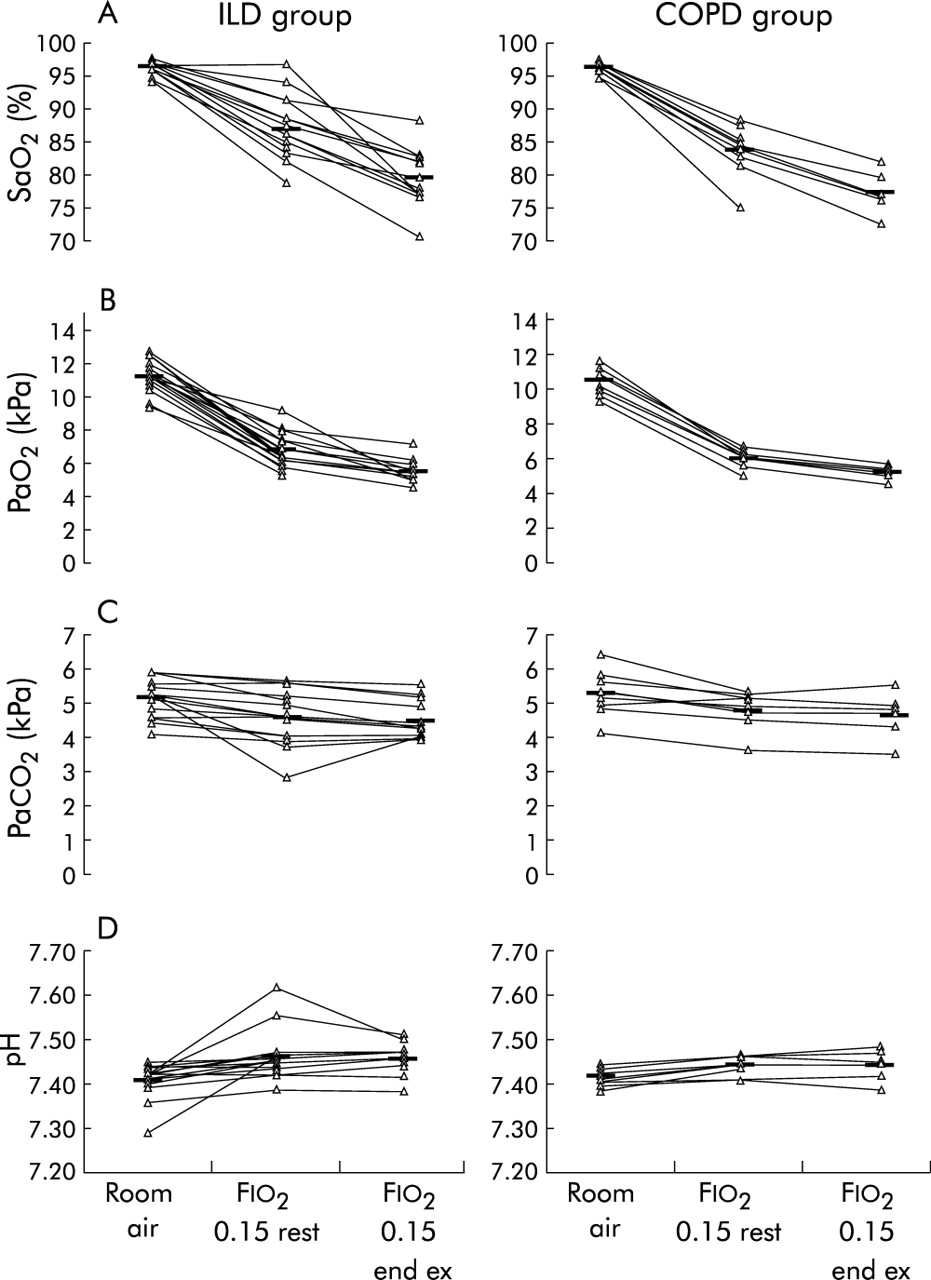
Individual arterial blood gas tensions and mean values are shown in fig 1. Mean (SD) values and statistical significance using paired or unpaired t tests as applicable are presented in table 2.
Comparison of mean (SD) arterial blood gas tensions between 15 patients with interstitial lung disease (ILD) and 10 with chronic obstructive pulmonary disease (COPD) at sea level at rest (room air), on 15% oxygen at rest for 20 minutes (Fio2 0.15 rest), and after a walking task on 15% oxygen (Fio2 0.15 end ex)

Comparison of arterial blood gas tensions between patients with interstitial lung disease (ILD) and those with chronic obstructive pulmonary disease (COPD) at sea level at rest (room air), on 15% oxygen at rest for 20 minutes (Fio2 0.15 rest), and after a walking task on 15% oxygen(Fio2 0.15 end ex). Individual and mean values of (A) arterial oxygen saturation (Sao2), (B) arterial oxygen pressure (Pao2), (C) arterial carbon dioxide pressure (Paco2), and (D) arterial pH.
The mean size (with 95% confidence intervals) of the changes in oxygenation are shown in table 3.
Mean size and 95% confidence intervals (95% CI) of changes in arterial oxygen saturation (Sao2) and arterial oxygen pressure (Pao2) between values at sea level at rest (room air), on 15% oxygen at rest for 20 minutes (Fio2 0.15 rest), and after a walking task on 15% oxygen (Fio2 0.15 end ex) in 15 subjects with interstitial lung disease (ILD) and 10 with chronic obstructive pulmonary disease (COPD)
Due to pre-determined test termination, if Spo2 fell below 80%, two ILD and one COPD subject did not complete 20 minutes resting Fio2 0.15. Only three COPD and five ILD subjects completed the 50 metre exercise task. Subjects who survived the Spo2 >80% criteria were plotted on a survival curve (fig 2). Individual test end points, lowest Sao2, and whether they had recently flown are shown in table 4.
Baseline arterial oxygen saturation (Sao2), Sao2 at end stage of inhaling 15% oxygen at rest (Fio2 0.15), extent of study reached, and Sao2 for each subject with interstitial lung disease (ILD) and chronic obstructive pulmonary disease (COPD)

Curve plotting subject survival by >80% saturation by pulse oximetry (Spo2) during a walking task on 15% oxygen (Fio2 0.15).
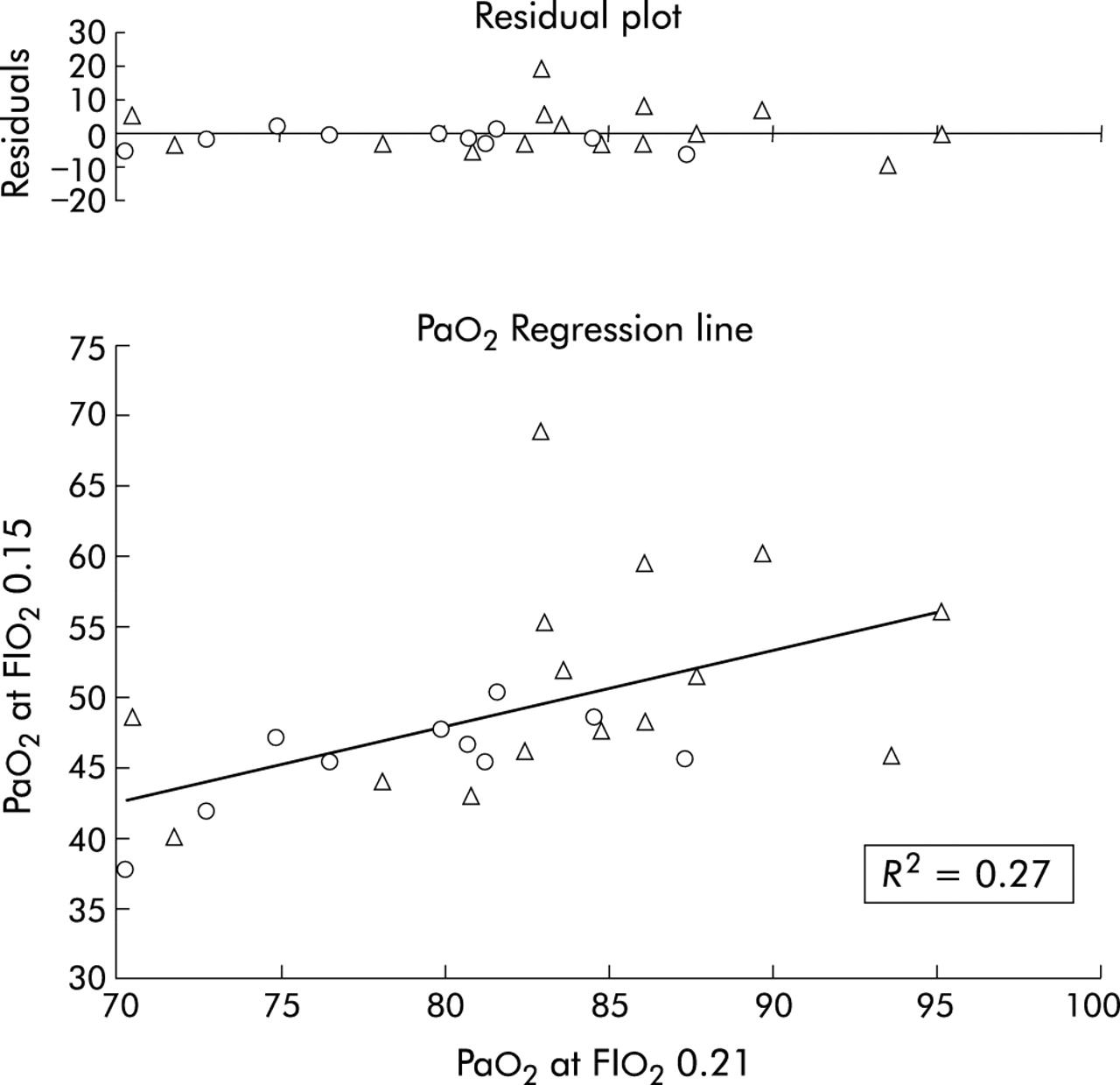
Linear regression analysis is presented in fig 3 with individual residuals plotted. From our data, the Pao2 at Fio2 0.15 can be calculated from a Pao2 collected at sea level (Fio2 0.21) according to the following formula:

Plot of arterial oxygen pressure (Pao2) at 15% oxygen at rest (Fio2 0.15) against Pao2) at sea level (Fio2 0.21) with residuals for individual subjects with interstitial lung disease (triangles) and chronic obstructive pulmonary disease (circles). Regression (solid line): Pao2 Fio2 0.15 = Pao2 Fio2 0.21 * 0.54 + 4.7.

Fifty two percent of our subjects had flown in the last 2 years, 80% in the last 5 years. Approximately half of the subjects reported drinking alcohol (48%), caffeinated drinks (64%), and visiting the lavatory (52%) during the flight. There were no reports of in-flight shortness of breath, headache, or dizziness, but there were two reports of “fatigue” attributed to air travel. Fifty four percent were planning to fly in the near future and 64% were unaware of “lower oxygen levels” on aircraft.
During “room air” and “Fio2 0.15 rest” there was no significant increase in Borg scale for COPD but there was for patients in the ILD group (p = 0.02).
Eighty eight percent of subjects passed all resting fit to fly guidelines with regard to Pao2, Spo2 or Sao2, and ambulatory status. The Pao2 of all subjects fell below 7.3 kPa and in 96% of cases it fell below 6.7 kPa while performing a mild exercise task on 15% oxygen. In 80% of subjects the Pao2 fell below 7.3 kPa at rest on 15% oxygen.
DISCUSSION
Predicting adverse consequences associated with hypoxia during a commercial flight is important. Patients with lung disease are concerned about health consequences and commercial airlines are concerned for their passengers, but also the economic costs and inconveniences associated with flight diversions. The results suggest that the degree of hypoxaemia under simulated cabin conditions cannot be predicted with confidence from resting measures. Furthermore, some subjects who under current guidelines do not warrant simulation testing, experience marked hypoxaemia when this is performed. Finally, some subjects who have flown without adverse effects can be shown to become markedly hypoxic during a simulation.
The data from subjects with COPD are in keeping with earlier studies which found that patients with COPD and mean Pao2 above 9.3 kPa at sea level develop mean values that fall below 7.3 kPa when exposed to normobaric hypoxia or hypobaria comparable to 8000 ft.5,10,18,19 When a minimal exercise challenge was added—either an incremental bicycle task10 or a stepping task5—there was a further fall in Pao2 of 1–1.5 kPa. None of these studies states whether the subjects had flown with or without adverse event. Our primary reason for inclusion of this group was to have a reference for our simulation test to this evidence base and to the existing guidelines.
A recent study has studied the effect of hypobaric hypoxia on patients with restrictive lung disease.20 However, while this study included some subjects with ILD, it also included others with a variety of other forms of restrictive lung disease. It is therefore difficult to establish direct comparability with our data. For all that, the results showed a similar response to this study at resting hypobaria equivalent to 8000 ft (mean 6.5 kPa) with a further fall to 5.1 kPa after a cycle task.
On standard measures of disease severity, the COPD group had more severe disease. We did not seek to recruit cohorts with similar severity as these are essentially incomparable disease processes. It can be said that the gas exchange abnormality, as assessed by measurement of carbon monoxide transfer factor, was similar. Resting blood gas tensions and the frequency and extent of hypoxia were similar for COPD and ILD subjects, but the COPD group experienced greater desaturation while breathing the hypoxic gas at rest.
Most of our subjects passed current resting fitness to fly guidelines.6–9 Only three subjects would have warranted further investigation by one guideline due to borderline pulse oximetry measures with an additional risk factor.8 However, the majority of subjects proceeded to “fail” the altitude simulation test at rest and almost all the remainder fell below an acceptable level of oxygenation when a minor exercise task was added. In-flight emergencies are rare; COPD is common. It is therefore reasonable to suggest that many similar patients fly now and do so without acute adverse events. We therefore do not advocate wider screening of patients who have ILD or COPD with an altitude simulation test until treatment decisions based on the simulation test can be better validated. More and better information on the clinical characteristics of patients with lung disease who do experience problems during flight is required. As these events are rare, this may be best achieved with a central register that would then be analogous to some of the rare congenital malformation databases. It would be in the interests of airlines to provide relevant information in relation to the cabin air conditions, particularly pressurisation schedules on the flight in question.
Approaching the problem from the other end, prospective evaluation of a large number of patients with lung disease who plan to fly is also required. This evaluation could be simple—limited to diagnosis and resting blood gas estimation. It should be correlated with the travel itinerary and any subsequent adverse events experienced. Altitude simulation studies are time consuming and only a subset of such patients could have a formal altitude simulation test. Significant clinical and ethical issues may well arise in patients who volunteer for such a study.
After careful consideration in conjunction with our institutional ethics committee, we resolved not to inform subjects in this study of their individual test results. To do so might have generated concern about the risks of flying when the magnitude of that risk was uncertain. Furthermore, there may be implications for travel insurance. In this study the limits of this “no tell” policy were not tested by what might be considered extreme hypoxia (Pao2 below 6.7 kPa in any subject). Presumably, any clinical centre would have to define policies for dealing with research subjects and the parameters outside which more complex evaluation or in-flight oxygen was recommended.
In summary, hypoxaemia considered severe enough to put a patient with ILD at risk during a commercial air flight cannot be accurately predicted from resting blood gas determination at rest. This exposes some limitation in the evidence base used to inform patients with lung disease of the potential hazards of commercial flights. While further data are accrued, we recommend that the existing guidelines be followed in relation to prediction and minimisation of these risks.