Article Text

Abstract
Background: Acid induced pneumonitis resulting in acute respiratory distress syndrome (ARDS) is characterised by increased alveolar permeability and accumulation of neutrophils. It is hypothesised that vascular endothelial growth factor (VEGF) is involved in the development of lung oedema. Furthermore, lower levels of VEGF are detected in bronchoalveolar lavage fluid from patients with ARDS than from non-ARDS patients. We hypothesised that VEGF acts cytoprotectively and have investigated this possibility in vitro with A549 cells.
Methods: A549 cells were incubated in 24 well culture dishes 24 hours before exposure to acid, then incubated with serum free medium containing various concentrations of HCl for 30 minutes at 37°C in 5% CO2. The acidified medium was changed to normal complete medium; at specified incubation periods the supernatants were collected and the VEGF concentration measured and the number of adherent cells counted.
Results: Proliferation of A549 cells and VEGF production were suppressed for at least 48 hours in HCl at a concentration of 50 mM. Restoration of cellular proliferation occurred following exogenous administration of VEGF (concentration of 1–250 ng/ml) and was inhibited by co-incubation with neutralising anti-VEGF antibody, indicating an interaction between VEGF molecules and A549 cells. Control cells were not influenced by administration of exogenous VEGF or anti-VEGF antibody. Treatment with neutralising anti-VEGF receptor (VEGFR) antibodies against VEGFR-1 and VEGFR-2 suppressed proliferation of acid exposed A549 cells but had no effect on control cells.
Conclusions: Exogenous VEGF interacts with VEGFR-1 and VEGFR-2 on the surface and regulates the proliferation of injured alveolar lining epithelial cells in an autocrine or paracrine fashion.
- VEGF
- acid induced lung injury
- cytoprotection
Statistics from Altmetric.com
Acid induced or aspiration pneumonitis is defined as acute lung injury following aspiration of regurgitated gastric juice or gastric contents.1 This syndrome occurs typically in patients with disturbed consciousness resulting from drug overdose, seizures, a massive cerebrovascular accident, or the use of anaesthesia. A pH of <2.5 and a volume of gastric aspirate of >0.3 ml/kg body weight (20–25 ml in adults) are required for the development of acid induced pneumonitis. A biphasic pattern of lung injury occurs following aspiration of gastric juice. The first phase, which reaches its peak 1–2 hours after aspiration, is the increased permeability of the alveolar-capillary interface. The second phase peaks at 4–6 hours following aspiration and is associated with infiltration of neutrophils into the alveoli and lung interstitium. Although many studies have been performed to confirm that the infiltrated neutrophils play the main role in the second phase of acid induced lung injury,2–6 the first phase has not been satisfactorily analysed.
Vascular endothelial growth factor (VEGF) is an endothelial cell specific mitogen that induces neovascularisation during normal organ development and various pathological processes.7 It has been speculated that VEGF plays a role in the generation of lung oedema—the first phase of ARDS—because (1) it is a vascular permeability factor, and (2) it is constitutively synthesised by human bronchial and alveolar epithelial cells and can be further induced by stimuli such as transforming growth factor (TGF)-β or hypoxia.8–10 However, it has been reported that the concentrations of VEGF in bronchoalveolar lavage (BAL) fluid of patients with ARDS are significantly lower than those of normal volunteers or patients with non-ARDS disease.11,12 Although the role of VEGF in the airways of normal lung is unknown, we hypothesised that VEGF produced by alveolar lining cells maintains the alveolar tissue milieu. We have investigated VEGF secretion in acid exposed alveolar lining model A549 cells in vitro.
METHODS
Cell culture
A549 alveolar epithelial-like cells were cultured in F-12K nutrient mixture medium (Kaighn's modification, Life Technologies, NY, USA) supplemented with 10% fetal calf serum, 50 U/ml penicillin, and 50 μg/ml streptomycin (complete medium) and incubated at 37°C in 5% CO2.
Acid exposure
Specified cell densities (5 × 10⁴, 1 × 10⁵, or 2 × 10⁵ cells) were placed in each well of a 24 well culture dish. After incubation for 24 hours, the medium was exchanged for serum free medium containing 10 mM, 50 mM, or 100 mM HCl. Control cells were exposed to sterile distilled water in the same volume as in the study group with 100 mM HCl. After incubation for 30 minutes at 37°C in 5% CO2, the acidified medium was discarded and the cells were washed several times with serum supplemented complete medium. Since we had previously confirmed that the pH of the culture medium after replacement of the acidified medium ranged between 7.2 and 7.4, the residual or carry over of HCl in these experiments was negligible. The pH of the acidified medium with 10 mM, 50 mM, and 100 mM HCl after incubation for 30 minutes in 5% CO2 was 7.3, 2.2, 1.1, respectively. The cells were incubated in 1 ml complete medium for specified time periods (3, 6, 12, 24, 36, 48 hours) and the supernatants were then collected and stored at –70°C until measurement of VEGF. The adhesive cells were treated with trypsin-EDTA (Life Technologies) and the number of cells counted with a haemacytometer. Counting was performed in at least four wells for each experimental condition.
Measurement of VEGF
Concentrations of human 165- and 121-amino acid isoforms of VEGF (VEGF165, VEGF120) were determined with the Quantikine VEGF Immunoassay (R&D Systems Inc, Minneapolis, MN, USA) according to the manufacturer's instructions.
In vitro administration of exogenous human VEGF
The proliferation of acid exposed A549 cells after administration of exogenous human VEGF was investigated. Before exposure to acid, A549 cells were seeded and cultured at a cell density of 1 × 10⁵ cells per well in a 24 well culture dish for 24 hours. After exposure to 50 mM HCl for 30 minutes the cells were cultured further with complete medium containing recombinant human VEGF165 (Genzyme-Techne, Minneapolis, MN, USA) at various concentrations (1, 10, 25, 100, 250 ng/ml) for 24 hours. The adhesive cells were detached by trypsin-EDTA and the number of cells was counted in at least six wells for each experimental condition.
Neutralisation by anti-VEGF antibody
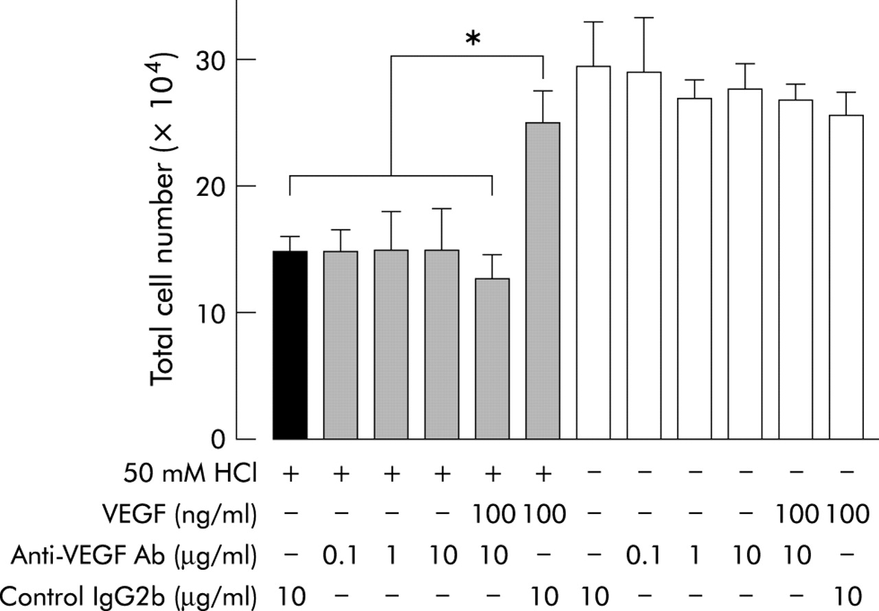
To evaluate whether VEGF regulates the recovery of proliferation of A549 cells exposed to acid, acid exposed cells were co-incubated simultaneously with exogenous VEGF and neutralising anti-VEGF antibody. Before acid exposure A549 cells were seeded at 1 × 10⁵ cells per well in a 24 well culture dish. After 30 minutes exposure to 50 mM HCl, the cells were incubated with 1 ml complete medium containing 100 ng/ml exogenous VEGF165 and the neutralising monoclonal anti-human VEGF165 antibody V4758 (Sigma, St Louis, MO, USA) at a concentration of 1 or 10 μg/ml for 24 hours.13 In control experiments class matched mouse IgG2b (Sigma) was added at a concentration of 10 μg/ml instead of anti-VEGF165 antibody. The number of adhesive cells was then counted after treatment with trypsin-EDTA in at least six wells for each experimental condition.
Treatment with anti-VEGFR antibodies
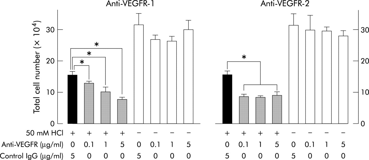
To determine whether the proliferation of A549 cells is mediated by VEGF receptor(s) on the surface of the cells, A549 cells at a density of 1 × 10⁵ cells per well in a 24 well dish were exposed to 50 mM HCl for 30 minutes and then incubated with 1 ml complete medium containing the neutralising monoclonal anti-human VEGFR-1 or VEGFR-2 antibody (R&D Systems Inc) at concentrations of 0.1, 1 or 5 μg/ml for 24 hours. These antibodies target the extracellular domains of VEGFR-1 and VEGFR-2, respectively. Class matched mouse IgG (Sigma) was added at a concentration of 5 μg/ml instead of anti-VEGFR-1 or anti-VEGFR-2 antibody in control experiments. The number of adhesive cells was counted after treatment with trypsin-EDTA in at least four wells for each experimental condition.
Data analysis
Data are presented as mean (SD). Statistical analysis was performed with ANOVA using Statview J 5.0 (Abacus Concepts Inc, Berkeley, CA, USA). A p value of <0.01 was considered significant.
RESULTS
Proliferation of A549 cells after acid exposure
The number of adhesive cells was significantly decreased after exposure to 50 mM and 100 mM HCl in the specified cell densities (5 × 10⁴, 1× 10⁵, 2 × 10⁵ per well) for each incubation time period (p<0.005 for each comparison, fig 1). In contrast, the cell numbers of those exposed to 10 mM HCl were not significantly different from the negative control cells (p>0.1 for each comparison). We had previously determined whether acid exposure increased dead or floating A549 cells by counting the number of adhesive cells after exposure to acid for 30 minutes. No increase in the number of dead or floating cells was seen after acid exposure at concentrations of 10, 50, and 100 mM (data not shown).

Suppression of proliferation of acid exposed A549 cells. A549 cells at densities of (A) 5 × 10⁴, (B) 1 × 10⁵, or (C) 2 × 10⁵ cells per well in a 24 well culture dish were exposed to 10, 50, and 100 mM HCl for 30 minutes and incubated further at specified time periods. The cell numbers at 10 mM HCl concentration were not significantly different from those of the negative control cells (DW) in each cell density. * indicates that the number of cells treated with 50 mM or 100 mM HCl was significantly lower than those of cells treated with DW (p<0.005 for each comparison at each indicated time).
VEGF levels in the supernatants of acid exposed A549 cells
A549 cells at a cell density of 1× 10⁵ per well were exposed to various concentrations of HCl. The supernatant was collected at specified incubation time periods (3, 6, 12, 24, 36, 48 hours) for determination of VEGF concentration. The concentration of VEGF was almost zero following exposure to 50 mM and 100 mM HCl, and suppression of VEGF production persisted for at least 48 hours (table 1). The concentration of VEGF in the supernatants of cells exposed to 10 mM HCl was not significantly different from that of the negative control cells (p>0.05 at each incubation time period). The combined results in fig 1 and table 1 show that the production of VEGF per cell was significantly suppressed after exposure to HCl for 30 minutes at a concentration of ≥50 mM. The production of VEGF in A549 cells was significantly suppressed after exposure to acid with pH <2.5.
VEGF concentrations (ng/ml) in supernatants of A549 cells with acid exposure
In vitro administration of exogenous VEGF
The mean (SD) numbers of acid exposed cells co-cultured with exogenous VEGF were 14.1 (1.9) × 104 for 1 ng/ml, 14 (2.6) × 104 for 10 ng/ml, 14.1 (1.8) × 104 for 25 ng/ml, 21 (1.6) × 104 for 100 ng/ml, and 19.6 (2.4) × 104 for 250 ng/ml (fig 2). The numbers of cells exposed to 50 mM HCl were 9.2 (1.9) × 104. The numbers of acid exposed cells treated with exogenous VEGF at concentrations of 1–250 ng/ml were significantly higher than those of acid exposed cells without exogenous VEGF (p<0.004 for each comparison). In addition, cells treated with 100 ng/ml and 250 ng/ml VEGF were increased to a greater extent than those treated with 1 ng/ml, 10 ng/ml, and 25 ng/ml VEGF (p<0.035 for each comparison). These results indicate that restoration of the proliferation of acid exposed cells is dependent on the concentration of exogenous VEGF.

Administration of exogenous VEGF to acid exposed cells. A549 cells at a cell density of 1 × 10⁵ per well in a 24 well culture dish were exposed to 50 mM HCl and incubated in complete medium supplemented with exogenous VEGF for 24 hours. The numbers of acid exposed A549 cells co-cultured with various concentrations of exogenous VEGF (shaded columns) were higher than those of acid exposed cells (solid column). The open column indicates the cell number of negative control cells. * indicates that the number of cells treated with exogenous VEGF at 1 ng/ml, 10 ng/ml, and 25 ng/ml was higher than those of acid exposed cells without exogenous VEGF (p<0.004 for each comparison). ** indicates that the number of cells treated with exogenous VEGF at 100 ng/ml and 250 ng/ml was higher than those treated at 1 ng/ml, 10 ng/ml, and 25 ng/ml (p<0.035 for each comparison).
Neutralisation by anti-VEGF antibody
To evaluate whether VEGF molecules mediate the restoration of proliferation of acid exposed A549 cells, neutralising anti-VEGF antibody was administered to the culture medium after acid exposure (fig 3). The numbers of acid exposed cells incubated with anti-VEGF antibody were 14.5 (1.9) × 104 with 0.1 μg/ml anti-VEGF antibody, 14.7 (3.2) × 104 with 1 μg/ml anti-VEGF antibody, and 14.7 (3.5) × 104 with 10 μg/ml anti-VEGF antibody. These values were close to that of acid exposure alone (14.5 (1.3) × 104) (p>0.92 for each comparison). However, the number of acid exposed cells treated with 100 ng/ml exogenous VEGF was 25.0 (2.5) × 104, which was significantly higher than the number of acid exposed cells treated with either 10 μg/ml anti-VEGF antibody or 100 ng/ml exogenous VEGF (12.5 (2.0) × 104, p<0.003). Exogenous VEGF, anti-VEGF antibody, and normal mouse IgG2b did not influence the proliferation of the control A549 cells (open bars in fig 3). These results suggest that restoration of the proliferation of acid exposed A549 cells is regulated by a VEGF molecular interaction via binding with VEGF receptor(s).

Neutralisation by anti-VEGF antibody. Acid exposed A549 cells at a cell density of 1 × 10⁵ per well in a 24 well culture dish were incubated in complete medium supplemented with VEGF and anti-VEGF antibody for 24 hours. Exogenous VEGF, anti-VEGF antibody, and normal IgG2b did not influence the proliferation of control cells (open columns). The number of cells incubated with anti-VEGF antibody at 0.1, 1.0 or 10 μg/ml or cells treated with anti-VEGF antibody (10 μg/ml) and exogenous VEGF (100 ng/ml) was not significantly different from those of acid exposed cells (solid column) (p>0.13 for each comparison). * indicates that the number of cells treated with class matched control IgG2b and exogenous VEGF (100 ng/ml) was significantly higher than those of acid exposed cells or acid exposed cells treated with anti-VEGF antibody (p<0.005 for each comparison).
Treatment with anti-VEGFR antibody
Although we investigated the expression of VEGF receptors on A549 cells by western blotting and flow cytometric analysis, neither VEGFR-1 nor VEGFR-2 was detectable in our study (data not shown). In flow cytometric analyses, acid exposed cells and control cells were incubated with 10 mM EDTA in PBS. The cells were detached with a scraper and the expression of VEGF receptor molecules was analysed. We avoided damaging the molecules by the use of trypsin. From these results, the expression of VEGFR-1 and VEGFR-2 was found to be low on both injured and control A549 cells. To determine the presence of VEGF receptors on A549 cells functionally we co-incubated acid exposed cells with various concentrations of neutralising anti-VEGFR antibodies and found that both neutralising anti-VEGFR-1 and VEGFR-2 antibodies suppressed the proliferation of acid exposed A549 cells even at a concentration of 0.1 μg/ml for each antibody investigated (fig 4). Interestingly, this suppression by anti-VEGFR antibodies was not observed in the control A549 cells not exposed to acid (fig 4).

Treatment with neutralising anti-VEGFR antibodies. Acid exposed A549 cells at a cell density of 1 × 10⁵ per well in a 24 well culture dish were incubated in complete medium supplemented with anti-VEGFR antibodies for 24 hours. Anti-VEGFR-1 and anti-VEGFR-2 antibodies did not influence the proliferation of control cells (open columns). * indicates that the number of acid exposed cells (shaded columns) incubated with anti-VEGFR-1 and anti-VEGFR-2 antibodies at 0.1, 1.0 or 10 μg/ml decreased significantly compared with those of acid exposed cells treated with class matched control IgG (solid columns; p<0.0024 for each comparison). Following treatment with anti-VEGFR-1 antibody the number of acid exposed cells was decreased as opposed to the increase in concentration of the antibody. This was not observed in acid exposed cells treated with anti-VEGFR-2 antibody.
DISCUSSION
Acid induced or aspiration pneumonitis is recognised as a model of ARDS characterised by an increase in alveolar permeability and lung oedema caused by acute diffuse injury of the alveolar capillary wall.1 The concentrations of VEGF in BAL fluid were analysed as it is thought that VEGF is involved in the pathogenesis of ARDS.7–9 However, the VEGF levels in BAL fluid are lower in patients with ARDS than in non-ARDS patients.10,11 For example, Maitre et al10 found VEGF levels in BAL fluid of 14.3 (11.1) pg/ml in ARDS patients and 76.8 (51.1) pg/ml in non-ARDS patients. These results indicate that increased alveolar permeability is not induced by increased VEGF levels in the alveolar space.
In our study acid exposure (pH <2.5) suppressed the production of VEGF and the proliferation of A549 cells. Furthermore, exogenous VEGF restored the proliferation of acid exposed A549 cells. Interestingly, this phenomenon was not observed in cells not exposed to acid. Treatment with neutralising anti-VEGF antibody showed that restoration of the proliferation of acid exposed cells was regulated by the ligand receptor interaction of the VEGF molecule. Two high affinity transmembrane tyrosine kinase receptors for VEGF have been described—fms-like tyrosine kinase-1 (Flt-1 or VEGFR-1)14 in which the expression appears to be restricted to the endothelial cells, and kinase insert domain containing receptor/fetal liver kinase-1 (KDR/Flk-1 or VEGFR-2).15 We were unable to demonstrate the presence of VEGF receptors on A549 cells by western blot or flow cytometric analyses so we evaluated the presence of VEGF receptors on A549 cells functionally by treatment with neutralising anti-VEGFR antibodies. Interestingly, the proliferation of A549 cells was suppressed by neutralising antibodies against both VEGFR-1 and VEGFR-2 after acid exposure, but this suppression was not observed in the control cells (fig 4). Although we investigated whether apoptotic cells were increased after acid exposure or after treatment with neutralising antibodies (anti-VEGF antibody, anti-VEGFR-1 antibody, or anti-VEGFR-2 antibody) by histological evaluation of pycnotic nuclei stained with 4,6-diamidino-2-phenylindole (DAPI, 1 μg/ml), there was no increase in the number of apoptotic cells (unpublished data). The results from the experiments with neutralising antibody indicate that the proliferation of injured A549 cells depends on the VEGF pathway after acid exposure. In contrast, control A549 cells are less dependent on VEGF to proliferate. Although we have shown that both anti-VEGF and anti-VEGF receptor antibodies block the restoration of the proliferation of A549 cells, we cannot completely dismiss the possibility of the release of other growth factors by VEGF.
VEGF is known to be important in maintaining the pulmonary circulation. Kasahara et al16 showed that capillary endothelial cells in the alveolar septa express both VEGF and VEGFR-2 to generate an autocrine loop and that the expression of both molecules decreased in emphysematous lung tissue. Boussat et al8 pointed out that human bronchial and alveolar epithelial cells constitutively synthesise VEGF, indicating that secreted VEGF in the bronchoalveolar space is not harmful to bronchial epithelial cells so is unlikely to induce pulmonary oedema.
Kaner et al17 showed that intratracheal administration of adenovirus vector that expresses VEGF at 109 plaque forming units in mouse resulted in pulmonary oedema. It was not possible to compare their results with ours because of the different methods used to introduce VEGF (we administered VEGF exogenously while they administered it endogenously into bronchial epithelial cells using an adenovirus vector). The syngeneic action of VEGF and the virus may have resulted in increased permeability of the alveolar-capillary interface.18 It is possible that pulmonary oedema is generated by exogenous VEGF, but our data indicate that VEGF protects injured alveolar lining cells even at a minimal dose of 1 ng/ml. Low dose VEGF may therefore provide a new approach to the treatment of acid induced pneumonitis and ARDS.
Acknowledgments
This work was partially supported by grants from Juntendo University, grants-in-aid for scientific research from the Ministry of Education, Culture, Sports, Science, and Technology of Japan (14570562) and grants-in-aid for scientific research on priority areas from the Ministry of Education, Culture, Sports, Science, and Technology of Japan (1402114).