


Abstract
The symptom burden resulting from sleep-disordered breathing (SDB) in patients with mild-to-moderate congestive heart failure (CHF) is unclear. The current authors monitored 24-h activity levels and compared subjective and objective measures of daytime sleepiness in 39 CHF patients, New York Heart Association class 2–3, on optimal medication. A total of 22 patients were classified as SDB (apnoea/hypopnoea index (AHI) median (range) 22.3 (16.6–100) events·h−1), and 17 as no SDB (NoSDB; AHI 3.7 (0–12.3) events·h−1). SDB was defined as AHI ≥15 events·h−1. Patients were assessed by 24-h activity monitoring (actigraphy) for a period of up to 14 days, a single objective sleepiness test (Oxford Sleep Resistance test) and Epworth Sleepiness Scale.
The duration of daytime activity was significantly shorter in the SDB group compared with the NoSDB group. The SDB group also had increased time in bed and poorer sleep quality, as shown by the fragmentation index. Objectively the SDB group when compared with the NoSDB group were significantly sleepier, subjectively the groups did not differ. The amount of napping was similar for both groups.
Despite the lack of subjective symptoms of daytime sleepiness, congestive heart failure patients with sleep-disordered breathing were objectively sleepier during the day and had reduced daytime activity with longer periods in bed and poorer sleep quality when compared with those without sleep-disordered breathing.
Excessive daytime sleepiness is a key feature of obstructive sleep apnoea (OSA). Repetitive arousals from sleep occur at the termination of an apnoea and result in sleep fragmentation with symptoms of sleepiness. This can lead to a reduced quality of life, occupational problems and an increased risk of road traffic accidents 1. Treatment of OSA with continuous positive airway pressure (CPAP) therapy is strongly linked to the relief of excessive daytime sleepiness 2, 3.
Sleep-disordered breathing (SDB) is common in patients with severe congestive heart failure (CHF) 4, 5. However, unlike patients with OSA, CHF patients with SDB frequently do not report symptoms of excessive daytime sleepiness, despite having a high apnoea/hypopnoea index (AHI) 6. Nevertheless, CHF patients with SDB are objectively sleepy during the day, regardless of not reporting subjective symptoms 6. Further objective measures of daytime sleepiness may help with assessing the symptom profile of these patients.
Actigraphy is a tool which has been widely used in sleep research to monitor activity over 24-h periods 7, 8 in a variety of patient groups 9, 10. Actigraphy has been shown to correlate well with polysomnography 7, 11. It can be used to define wake epochs during nocturnal sleep, and has also been used to monitor daytime napping in healthy populations 8. Daytime napping and 24-h activity levels have not previously been documented in CHF patients with or without SDB. The purpose of the present study was to monitor 24-h activity levels in CHF patients with SDB over a 2-week period using actigraphy. More specifically, the current authors compared 24-h activity levels between CHF patients, with or without SDB. In addition, subjective and objective measures of daytime sleepiness were compared.
The current authors hypothesised that CHF patients with SDB would have decreased levels of daytime activity, compared with CHF patients with no SDB (NoSDB), which may in turn explain the lack of excessive daytime sleepiness.
METHODS
Subjects
A group of 39 consecutive male CHF patients were recruited from cardiology clinics as part of a wider study assessing the prevalence of SDB in mild-to-moderate heart failure patients 12. All patients who had taken part in the prevalence study (n = 55), were approached and invited to participate in the present study, apart from those who had started to undergo treatment for SDB (n = 8). Eight patients declined to take part in the study. Selection was not determined by the presence or absence of daytime sleepiness.
Inclusion criteria involved all patients receiving optimal medical therapy (table 1⇓) and having stable heart failure, defined by no changes in cardiac medication 4 weeks prior to and during the study. Individuals with a forced expiratory volume in one second (FEV1)/forced vital capity (FVC) ratio of <0.70 or chronic neurological disease, were excluded. Patients being treated for sleep disorders or with concurrent diseases that were likely to affect the control of breathing for other reasons, i.e. cerebral infarction, chronic neurological disease, or receiving hypnotic/sedative/opiate drug treatment, were also excluded. Patients were divided into two groups: SDB (n = 22) and NoSDB (n = 17). Systolic function was not significantly different between the two groups after grouping for SDB status. However, no patients were removed from the analysis on the basis of matching for systolic function, and the matching of the groups occurred by chance.
Group characteristics
Sleep staging was scored using the standard methods of Rechstaffen and Kales [13] for 30-s epochs. SDB was measured by polysomnography and defined as an AHI >15 events·h−1 4. Respiratory events were scored as follows: apnoea, the presence of no airflow for >10 s; hypopnoea, a 50% reduction in airflow for >10 s in the presence of a 4% desaturation or an arousal; central sleep apnoea (CSA), ≥50% of apnoeas central or mixed; OSA, >50% of apnoeas obstructive. The mean±sd time between polysomnography and actigraphy was 3±3.3 months.
At the time of polysomnography, the patient's height and weight were measured and their body mass index (BMI) calculated (weight·height−2). A complete two-dimensional and Doppler echocardiography study was performed for each patient. Left-ventricular ejection fraction (LVEF) was assessed by single plane Simpson's method 14. Patients underwent ergometric cardiopulmonary exercise test (Oxycon; Jaeger Toennies Ltd, Höchberg, Germany) from which peak oxygen uptake (V′O2) was calculated. Carbon dioxide arterial tension (Pa,CO2) was measured from blood taken from the ear lobe, prior to polysomnography. All cardiac measurements were taken within 24 h of the overnight polysomnography.
All participants gave written, informed consent, and the study was approved by the local ethics committee.
Protocol
The patients wore an Actiwatch® (Model AW4; Cambridge Neurotechnology Ltd, Cambridge, UK) on their nondominant wrist for 2 weeks. Daily sleep diaries were kept to report bed and get-up times, nocturnal urination events and daytime napping. Individual daily data were omitted if the Actiwatch® was removed for >2 h. The current authors obtained >13 days of 24-h data from 28 of the 39 patients (72%) involved; 11 or 12 days' data was compiled in a further seven patients (18%) and 10, 9, 8 and 7 days' data were collected for each subject, respectively, in the remaining 10%. One subject failed to wear the Actiwatch® during the day on 12 days out of the 14 days studied, and one failed to wear the Actiwatch® throughout the night on all nights, consequently their daytime and nocturnal data, respectively, were removed.
Actigraphy measurement
The Actiwatch® measures activity with a piezo-electric accelerometer that records intensity, amount and duration of movement in all directions. All movement over 0.05g with a maximum sampling frequency of 32 Hz is recorded. Actigraphical data from 1-min epochs were collated and automatic scoring of sleep was performed using a validated algorithm 15. This algorithm modifies recorded activity counts in one epoch (1 min) according to the level of activity in the surrounding 2 min (±2 min) to give a final activity count for each epoch. The total value is used to decide if the epoch is scored as wake or sleep. During the current analysis, a threshold of >40 counts·epoch−1 was used to define wake.
Nocturnal activity analysis
For the nocturnal period, bed and get-up times were set according to cues (actigraph event marker or sleep diary entry). If the times recorded using the actigraphy event marker differed from the sleep diary then the actigraphy event marker was considered more reliable compared with the patient's estimations entered in the sleep diary. The onset and end of sleep were also manually estimated using actigraphy from the first and last periods of quiescence, respectively; however, this may or may not have reflected sleep and wake times that would have been defined had the changes in the frequency of the electroencephalogram been measured. Time in bed (TIB), sleep latency (SL), sleep efficiency (SE), movement time and fragmentation index (calculated as the percentage of the number of phases of immobility of ≤1 min against the total number of immobility phases; immobility phase: >1 consecutive epochs with a score of zero) were all calculated by the actigraphy sleep–wake algorithm.
Daytime activity analysis
The daytime period was analysed using actigraphy software following manual prompts. Activity was taken to be any movement associated with wakefulness; the actiwatch did not discriminate between arm movement activities and whole body exercise. The duration of daytime activity was defined as the time between the first period of activity following sleep and the last period of activity preceding sleep. Activity counts per epoch during the day were added to produce the total daytime-activity count per day.
Nap analysis
Naps were objectively analysed using the actigraphy analysis software and subjectively analysed using cues from patients' individual sleep diaries. The minimum nap period was set at 10 min. The algorithm for nap analysis classified an epoch as sleep if the activity counts in an epoch fell below a certain threshold; this was set at zero. For each daytime period an objective and subjective mean nap time was calculated.
Subjective and objective sleepiness assessment
An objective SL test (Oxford Sleep Resistance test (OSLER); Stowood Scientific Instruments, Oxford, UK) was carried out 16. In the majority of patients this was done at the time of the actigraphy study (n = 27); in the remaining patients (n = 12) this was assessed within 8 weeks (median 6, range 1–8 weeks). The OSLER test was undertaken between 13:30 h and 19:30 h (median was 16:30 h) in a darkened, quiet room. The patients had not eaten or had any caffeinated drinks within 1 h prior to the test. The patients were asked to maintain wakefulness for the maximum testing time of 40 min. Sleep was considered to have occurred when seven consecutive cues were missed, which corresponded to a time of 21 s. An Epworth Sleepiness Scale (ESS) questionnaire 17 was completed prior to the start of actigraphy.
Statistical analysis
All data were tested for normality. Unpaired t-tests or Mann–Whitney rank-sum tests were used to compare groups for variables and group characteristics. Z-tests were used to compare proportions. ANOVA was used to compare the time of day that the OSLER test was taken for all subjects. Results are expressed as mean±sd or median (range). Statistical significance was accepted as p≤0.05.
RESULTS
Subject characteristics
A total of 39 patients were studied. There were no significant differences in age, BMI, LVEF, peak V′O2, New York heart Association, Pa,CO2, hours of employment, nap opportunity or alcohol consumption between CHF patients with and without SDB. Group characteristics are shown in table 1⇑.
Polysomnography data
The SDB group had a median (range) AHI of 22.3 (16.6–100) and the NoSDB group 3.7 (0–12.3) events·h−1 (p<0.001). Of the SDB group, 12 patients had CSA and 10 patients had OSA. The SDB and NoSDB groups differed significantly in desaturation index (p<0.01), SE (p = 0.01) and arousal index (p<0.001). CHF patients with SDB tended to have more stage 1 sleep, although this did not reach statistical significance. Both groups had similar amounts of deep and rapid eye movement sleep. Polysomnography data are shown in table 2⇓.
Polysomnography data
24-h activity from the actigraph
The 24-h activity data, over 2 weeks, in one CHF patient with SDB and one without SDB are shown in figure 1⇓. Comparisons of each group's mean nocturnal parameters are given in table 3⇓. CHF patients with SDB spent a significantly longer TIB (p = 0.01) compared with the NoSDB group. However, their sleep was more disturbed, as shown by the significantly increased movement time (p = 0.006) and fragmentation index (p = 0.003). SE tended to be worse in the SDB group, although this did not reach statistical significance (p = 0.06). SL did not differ significantly between the two groups (p = 0.17).

Representative actigrams are >2 weeks (dual-day display) from congestive heart failure (CHF) patients with a) sleep-disordered breathing (SDB) and b) no SDB. Patients without SDB have a significantly more active daytime profile. Sleep fragmentation is similar in the two patients presented in this figure, despite being significantly increased in the group's mean for CHF patients with SDB. Notice that peaks of activity during the night are associated with nocturnal urination events.
Actigraphy data
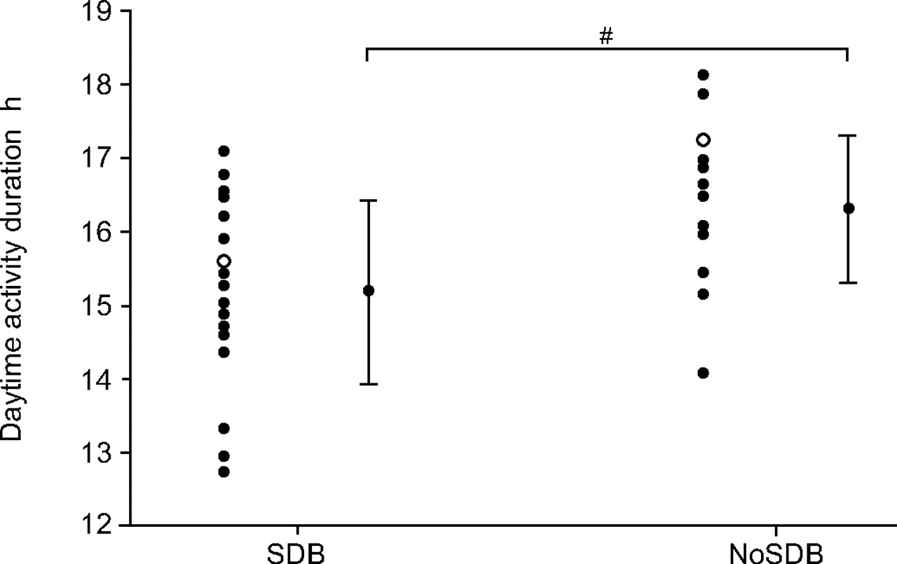
Daytime activity levels in one CHF patient with SDB and one without SDB are shown in figure 2⇓. The individual and group mean daytime activity are shown in figure 3⇓. For the SDB group the daytime activity duration was significantly shorter than that of the NoSDB group (p = 0.004). Within this period the CHF patients with SDB tended to be less active, although this did not reach statistical significance (total activity counts p = 0.06; average activity/epoch p = 0.08; table 3⇑).

Examples of average 24-h activity levels for a congestive heart failure (CHF) patient a) with no sleep-disordered breathing NoSDB and b) with SDB. These data highlight the longer time in bed and reduced activity in CHF patients with SDB. The traces also show daily nap times. Nap times are as reported in daily sleep diaries. The levels of activity do not reach the low levels seen during sleep as the plot is an average over a 2-week period, during which not all naps are at the same time or duration. Both patients had similar levels of sleep fragmentation; however, the intensity of movement (total activity counts) during sleep was greater in the CHF patient without SDB. This produced higher activity levels during the lights-out period.

The current authors correlated 24-h activity parameters with SDB severity on the basis of AHI. As a continuous variable, AHI weakly correlated with daytime activity duration (r2 = 0.15, p = 0.02). There were no further significant correlations between the parameters of Pa,CO2, LVEF and peak V′O2, which the current authors used to define cardiac status and the actigraphy measurements of TIB, SE, fragmentation index and daytime activity duration (p>0.05). These cardiac parameters also showed no correlation with AHI, ESS or OSLER test results (p>0.05).
There were no significant differences (p>0.05) in actigraphy results (TIB, SE, fragmentation index, daytime activity duration or total daytime activity counts) for those patients who had actigraphy at the time of polysomnography (SDB, n = 10; NoSDB, n = 6) and for those who had measurements at >1 month apart (SDB, n = 12; NoSDB, n = 11).
Subjective and objective daytime sleepiness
There was no significant difference between the SDB and NoSDB groups for subjective ESS (median (range); SDB: 7 (2–16); NoSDB: 9 (2–17); p = 0.55), yet objectively the SDB group were significantly sleepier on the OSLER test (SDB: 17.1 (3–40) min; NoSDB: 40.0 (12–40) min; p = 0.01).
OSLER results were further separated according to the time of day the test was taken (13:30–15:30, 15:31–17:30 and 17:31–19:30 h). The current authors found no significant differences in the SL recorded by the OSLER test for each time of day (p = 0.77).
Behavioural symptoms
The mean subjective total nap time per day was similar in the SDB and NoSDB groups; although there was a wide range of variability within each group (mean±sd; SDB: 22.9±17.9 min·day−1; NoSDB: 26.1±23.0 min·day−1; p = 0.69). Daytime napping was also analysed objectively from actigraphy; individual examples of naps are seen in figure 2⇑. The mean objective nap time was similar for the SDB and NoSDB groups (SDB: 26.1±27.2 min·day−1, NoSDB: 19.6±18.6 min·day−1, p = 0.36). There was no significant difference between the groups in the percentage of days when naps were taken (SDB: 64±48 % days, NoSDB: 49±30 % days; p = 0.26).
Nocturnal urination was reported at a higher frequency in the SDB group than the NoSDB group, although this did not reach statistical significance (SDB: 1.7±1.2 times·night−1, NoSDB: 1.0±0.9 times·night−1, p = 0.07).
DISCUSSION
This is the first study to monitor 24-h activity levels in CHF patients with and without SDB. The main findings were that CHF patients with SDB spent longer periods of time in bed, they had more fragmented sleep and tended to be less active during the day than a group of CHF patients without SDB, who had no significant differences in cardiac parameters, age or BMI. Consistent with previous studies, the current results show that CHF patients with SDB report very little daytime sleepiness, yet objectively there was a significant difference in daytime sleepiness. These data highlight the fact that CHF patients with SDB underestimate their daytime sleepiness symptoms. The current authors speculate that the reduced perception of sleepiness may, in part, be due to reduced levels of daytime activity with longer periods in bed.
The use of 24-h actigraphy, over a 2-week period, in the current study, allowed the authors to obtain accurate data on sleeping habits which cannot be determined during single, overnight polysomnography in a sleep laboratory. The increased sleep fragmentation on the actigraphy traces in the SDB group suggests that these patients are having disturbed sleep when at home. These data are consistent with the decreased SE and increased number of arousals scored in the polysomnography. Sleep fragmentation indices, from a number of different techniques, have been shown to be useful in identifying OSA patients with sleepiness, who are likely to respond to CPAP treatment 18. Thus, this could be a useful parameter in assessing which CHF patients with SDB would benefit from treatment.
The current authors found that subjective and objective measurements of sleepiness in CHF patients with SDB, produced different results. CHF patients with SDB are objectively, but not subjectively, sleepy during the day. This result has been seen in previous studies 6, 19. The ESS is widely and easily used in the clinical setting; however, the difference between subjective and objective sleepiness results within the CHF with SDB group could be accounted for as the ESS produces highly variable results 19, 20. The ESS may not always be applicable to older heart failure patients who no longer drive and have plenty of opportunity for daytime napping.
It has been speculated that the lack of differences in subjective sleepiness are due to overlapping symptoms of heart failure, including medications, with symptoms of SDB 4. An alternative explanation is that despite sleep fragmentation, CHF with SDB patients are able to maintain relatively normal levels of deep sleep. In the current study the percentage of deep sleep (11%) was comparable to that found in the Wisconsin Sleep Cohort study (n = 352; average being 9%); however, these data were collected in people who were younger (aged 30–60 yrs) than the current study population 21. It is known that increasing age decreases the amount of deep sleep 22, although reports differ as to the magnitude of the decrease. In one report, deep sleep in healthy males decreased from 19% in 16–25 yr olds, to 3% in 36–50 yr olds; with no further significant decreases in old age (71–83 yrs) 23. Alternatively, in the Sleep Heart Health Study (n = 2,685) deep sleep decreased from 11% in <54 yr olds, to 7% in 61–71 yr olds 24. Decreases in deep sleep can also occur in patients with respiratory disturbances, who are otherwise healthy 24. In patients with CHF and SDB, some 6, 25 but not all 4, 5, 26 studies have reported percentages of deep sleep that are comparable to those found in the present study.
The current authors investigated the levels of daytime napping in CHF patients. Both groups reported similar amounts of napping and these values closely matched the objective values recorded by actigraphy. This good reliability of nap reporting has not always been shown in actigraphy studies 27. In the recent study by Yoon et al. 8, older adults (mean age 66.2±4.9 yrs) were studied with actigraphy. Objectively they napped for 23.3 min·day−1, which correlates well with subjective and objective data in the current CHF patients. However, an earlier study reported that healthy, elderly subjects napped for 59.8 min·day−1 on average 28, which is significantly greater than either of the current patient groups. It is therefore difficult to say conclusively whether the levels of napping found in the present study are similar to a healthy, elderly population. There have been suggestions that nocturnal activity changes with increasing age 27 and that actigraphy algorithms are not necessarily equally accurate for adults at differing ages 29. However, since the current groups studied, with or without SDB, showed no significant differences in age and disease severity this contention is unlikely to have influenced the results obtained from the current study.
The present authors separated the data into two groups due to the symptoms of SDB being more common in CHF patients at an AHI of >15 events·h−1 4, despite some studies using an AHI >10 to define mild sleep apnoea 5, 30. An AHI of 15 is the value used by Javaheri et al. 4 in the benchmark prevalence study of 81 CHF patients to define SDB. Separation of the current data, using the same cut-off, allowed the current results to be compared with those of this seminal paper.
Limitations
A potential limitation of the current study was the variation in the time taken between polysomnography and actigraphy (3±3.3 months). Due to the unstable nature of CHF this may have led to small changes in cardiac parameters prior to actigraphy measurements. However, there were no changes in medication, any hospitalisations or decompensations during this time. Also, the number of patients in the SDB and NoSDB groups, who had polysomnography >1 month, apart from the actigraphy and OSLER test, was equal in both groups and there were no significant differences in all results between these sets of patients.
Due to the large amount of overlap between the current groups in activity levels, the present study would have been improved with a group of healthy elderly controls. The inclusion of healthy elderly controls could have highlighted that CHF alone reduces activity 31. However, from previous studies it is known that healthy elderly people (without CHF) have increased levels of daytime napping compared with healthy young controls 32, which may indicate that reduced daytime activity is a function of age rather than CHF. In this scenario there would continue to be a large amount of crossover between age-matched groups of CHF patients with and without SDB, but any small differences would highlight the impact of SDB on daytime activity levels.
The current authors used a single OSLER test. Mazza et al. 33 reported that a single 09:00 h OSLER test was as sensitive as three consecutive tests in identifying patients with significant daytime sleepiness. In the present study, patients were tested between 13:30 and 19:30 h (median: 16:30 h). This may have reduced the patient's ability to perform the test, due to their afternoon peak in physiological somnolence 33. However, the OSLER results were significantly different between CHF patients with and without SDB in terms of SL. The absolute values were similar to those of Pepperell et al. 6, who showed an improvement in objective daytime sleepiness, measured using four OSLER tests during 1 day, in a group of CHF patients with SDB, who were treated with adaptive servo controlled ventilation for a 1-month period. Furthermore, the current authors found no significant difference in the SL results of the OLSER test between those patients who had the test at different times during the afternoon.
The number of patients recruited was lower than the desired number calculated from a prospective power calculation. From the study by Rosenthal et al. 34, significant differences in daytime napping duration were found between severe (37.3±47.5 min·day−1) and mild (24.4±30.6 min·day−1) OSA patients. Using these differences, with a power of 80% and significance of 0.05, it was calculated that 90 patients would be needed in each group. Such high numbers of CHF patients with and without SDB were unavailable to the current authors over the study period selected; therefore, the maximum number of patients that agreed to partake in the current study were selected (see methods). There was no previous data available to perform a power calculation on activity levels. A retrospective power calculation (80% power; 0.05 significance) of the current daytime-activity duration data shows that groups of 13 would have produced significant results. However, 131 patients per group would have been required to find a significant difference in objective daytime napping.
Clinical implications
SDB in CHF is common and associated with sleep disruption, oxyhaemoglobin desaturation 4 and a poor prognosis 35, 36. It is therefore important to understand the full symptom profile of SDB in CHF patients in order to develop effective treatment regimes. Treatment of SDB in CHF patients with oxygen, CPAP, and adaptive servo-ventilation have reduced apnoeas by as much as 80% 25, 37, and 1 month of adaptive sero-ventilation has been shown to reduce objective daytime sleepiness, and produce a fall in brain natriuretic peptide and urinary metadrenaline excretion 6.
Patients with CHF have shown improved outcomes following exercise programmes 38. With further reduced activity levels in patients with SDB there may be further detrimental effects on the heart. Treatment of SDB may allow increased activity and an increased chance of cardiac improvement on an exercise programme.
Despite the lack of symptoms of daytime sleepiness, congestive heart failure patients with sleep-disordered breathing are objectively sleepier during the day, spend longer in bed, have more fragmented sleep and tend to have reduced daytime activity. This suggests that these patients may benefit from treatment. If treatment is to be evaluated effectively, subjective measures of sleepiness may not show significant improvements. However, actigraphy and the Oxford Sleep Resistance test may be useful in assessing improvements in sleep fragmentation, the length of time spent in bed and the objective sleepiness in these patients, and in assessing which patients may greater benefit from treatment.
Acknowledgments
The authors would like to thank M.R. Cowie, P.A. Poole-Wilson and H.F. McIntyre for allowing the recruitment of patients from their cardiology departments (National Heart and Lung Institute, Imperial College London and Royal Brompton Hospital, London, and The Conquest Hospital, Hastings, UK).
- Received May 31, 2005.
- Accepted December 12, 2005.
- © ERS Journals Ltd
References


