Article Text

Abstract
Compared with invasive ventilation, non-invasive ventilation (NIV) has two unique characteristics: the non-hermetic nature of the system and the fact that the ventilator-lung assembly cannot be considered as a single-compartment model because of the presence of variable resistance represented by the upper airway. When NIV is initiated, the ventilator settings are determined empirically based on a clinical evaluation and diurnal blood gas variations. However, NIV is predominantly applied during sleep. Consequently, to assess overnight patient–machine ‘agreement’ and efficacy of ventilation, more specific and sophisticated monitoring is needed. The effectiveness of NIV might therefore be more correctly assessed by sleep studies than by daytime assessment. The most available and simple monitoring can be done from flow and pressure curves from the mask or the ventilator circuit. Examination of these tracings can give useful information to evaluate if the settings chosen by the operator were the right ones for that patient. However, as NIV allows a large range of ventilatory parameters and settings, it is mandatory to have information about this to better understand patient–ventilator interaction. Ventilatory modality, mode of triggering, pressurisation slope, use or not of positive end expiratory pressure and type of exhalation as well as ventilator performances may all have physiological consequences. Leaks and upper airway resistance variations may, in turn, modify these patterns. This article discusses the equipment available for NIV, analyses the effect of different ventilator modes and settings and of exhalation and connecting circuits on ventilatory traces and gives the background necessary to understand their impact on nocturnal monitoring of NIV.
- Noninvasive ventilation
- ventilatory modalities
- bi-level positive airway pressure
- respiratory failure
- monitoring
- respiratory measurement
- sleep apnoea
Statistics from Altmetric.com
- Noninvasive ventilation
- ventilatory modalities
- bi-level positive airway pressure
- respiratory failure
- monitoring
- respiratory measurement
- sleep apnoea
Since the first studies in the early 1980s showing the usefulness of non-invasive ventilation (NIV) in the management of some forms of respiratory failure,1–4 the number of patients receiving this treatment both in the acute setting and at home is continuously increasing. This is explained by a growing number of indications in which the effectiveness of NIV has been proven, but also by major technological advances that led to the availability of high-performance portable ventilators as well as to the development of technical support infrastructure.5
When starting NIV, it seems desirable to verify whether this particular form of treatment is effective and has no untoward effects. Since home NIV is generally applied at night, nocturnal monitoring seems the best way to assess its effects. Although nocturnal monitoring of continuous positive airway pressure (CPAP) has been codified in the treatment of patients with obstructive sleep apnoea syndrome,6 this is not the case with NIV.7 8 Nocturnal monitoring of NIV is far more difficult and unforeseen problems arise for many reasons: (1) sleep can induce profound ventilatory changes, in particular in patients with respiratory insufficiency; (2) these ventilatory changes are also exacerbated by the interactions with a preset device—the ventilator can induce sleep disturbances by itself; (3) physicians caring for these patients may vary greatly in their methods of assessing the effects of NIV from a single blood gas measurement to full polysomnography.
The issue is complicated further because volume and pressure preset ventilators will respond differently in certain situations and because the type of mask used will also have different effects on monitoring parameters.9 The semiology of abnormal respiratory events during sleep is therefore completely different depending on the technology used. Pressure and flow dynamics also differ markedly among the different type of ventilators available for home care.
This article will deal with the equipment available for NIV, in particular the ventilator types, modes and settings. Its goal is to give the background necessary to understand their impact on nocturnal monitoring of NIV.
Issues of particular importance during NIV: leaks, upper airway resistance, type of exhalation port and NIV ventilators
Compared with invasive ventilation, NIV has two unique characteristics: the non-hermetic nature of the system that poses the potential risk of unintentional leaks and the fact that the ventilator-lung assembly cannot be considered as a single-compartment model because of the presence of a variable resistance represented by the upper airway (UA). Both situations may compromise the delivery of an effective tidal volume. As a consequence, increasing the delivered volume or the delivered inspiratory pressure during NIV does not necessarily result in increased effective ventilation reaching the lungs.10 When monitoring NIV using flow or pressure tracings, these two peculiarities will unavoidably influence the features of the respiratory traces.
Influence of unintentional leaks
Unintentional leaks are very common in NIV.11 12 Leakage may be absent or minimal when the patient is awake but may worsen during sleep as a result of the loss of voluntary control and decreased muscle tone. Leaks can take place at the mouth or between the skin and the mask, and air could also be deposited in the oropharyngeal reservoir and even flow into the digestive tract (‘internal’ leaks).13
Leaks can impair the quality of both ventilation and sleep. They can largely affect ventilator triggering, pressurisation, volume delivered, rate of inspiratory pressuring and cycling function and induce sleep fragmentation.
Influence of the upper airway
During NIV a variable resistance constituted by the UA is interposed between the ventilator and the lungs. The UA may change its resistance to airflow, compromising the delivery of an effective tidal volume to the lungs. Intermittent obstruction of the UA is common during NIV and may be related to two mechanisms. The first corresponds to obstructive events at the oropharyngeal level because of UA collapse as a result of insufficient expiratory airway pressure. This mechanism may be present in patients with an unstable UA.14 Another mechanism corresponds to episodes of intermittent obstruction at the glottic level reflecting cyclic glottic closure induced by hyperventilation, a type of ‘ventilation resistance’ reaction.15–18
Influence of type of exhalation device and connecting circuits
Whereas ICU ventilators classically use a double circuit with an integrated expiratory valve, two different types of circuits can be used to provide NIV. The first uses a similar assembly to those used in ICU devices and includes either single or double tubing in which inspiration and expiration are separated and a true expiratory valve is present so that carbon dioxide (CO2) rebreathing is not a significant problem (figure 1A). A second type does not have a true exhalation valve and often uses a single-limb circuit with a risk of rebreathing. To avoid rebreathing, this system includes a calibrated leak (called an intentional leak) either at the mask level or in the circuit (figure 1B). Single circuit pressure-targeted ventilators provided with a calibrated leak (called bilevel ventilators) are most commonly used for NIV nowadays. These devices cycle between a higher inspiratory positive airway pressure (IPAP) and a lower expiratory positive airway pressure (EPAP) that can be independently adjusted. With these devices, a minimum mandatory EPAP level of 4 cm H2O is needed to ensure an effective washout of CO2.20 Interestingly, a recent study showed that the exhalation port position influences CO2 rebreathing with a more efficient CO2 washout when the leak is positioned within the mask.21

Type of circuits used in non-invasive ventilation (NIV) with (A) an expiratory valve and (B) intentional leak, and typical traces obtained with these different assemblies. Note that when a circuit with an expiratory valve is used, the expiratory valve may be interposed in the circuit (single circuit) (A) or included in the ventilator (double circuit) (B); when a single circuit with intentional leak is used, the leak may be interposed in the circuit (C) or incorporated at the mask (D). Flow traces can be influenced by the type of circuit used, but also by the position of the pneumotachograph with regard to the expiratory device. In the case of a double circuit provided by an expiratory valve, the expiratory slope will reflect the expired volume only when the flow sensor is interposed between the mask and the exhalation device (trace 2 in A), but not when it is placed distally to the expiratory valve (trace 1 in A). On the other hand, when using a single circuit with intentional leak, the expiratory slope does not reflect the expired tidal volume and may be absent (B). In this case, the position of the pneumotachograph with regard to the leak device does not significantly change the expiratory slope. Modified from Perrin et al19 with permission.
When monitoring NIV using polygraphy, the mode of exhalation used and the position of the flow sensor (in relation to the expiratory device) both have a major influence on features of respiratory traces. Typical traces of these different assemblies can be seen in figure 1A and B.
Influence of the ventilator: ICU vs home devices
Both ICU and home ventilators can be used to deliver NIV. The main technical characteristic differentiating them is that, for the former, the driving pressure is supplied by compressed gas, whereas the latter devices incorporate their own pressure source. Nevertheless, as the type of ventilatory support that they provide is similar, this difference has little influence on respiratory traces. Therefore, when monitoring NIV, respiratory traces will have similar features whatever the type of positive pressure device used for ventilation.
Ventilator modes and settings: what's in a name?
When NIV was introduced there were a very limited number of modalities and types of ventilators with very few possible settings. We now have more than 30 brands offering numerous options for settings.22 Moreover, ventilators are not submitted to stringent medical regulations. This leaves manufacturers free to give different names to the same ventilator modalities and settings and even to ‘create’ new modalities that frequently correspond only to small modifications of a known class. This explains the wide variety of existing terminology describing NIV modalities.
Influence of the ventilator mode
Theoretically, NIV could be delivered using all the modalities used for invasive ventilation but, in real life, most ventilators used for NIV deliver either volume or pressure-targeted ventilation. Because the place of other anecdotal modalities proposed by some NIV devices such as synchronised intermittent mandatory ventilation or other ‘hybrid’ modalities is not yet clear, this paper will mainly focus on the former two modalities.
Volume-targeted ventilation (VTV)
In this modality the ventilator delivers a fixed volume during a given time and will generate whatever pressure is necessary to achieve this, regardless of the patient contribution to ventilation. Pressure in the airways (Paw) is not constant and results from the interaction between ventilator settings, compliance and resistance of the respiratory system and spontaneous inspiratory efforts (figure 2). It should be emphasised that any additional inspiratory effort will not lead to changes in delivered volumes or flows but only result in a decrease in Paw. As each breath is delivered with the same predetermined flow-time profile and as the area under the flow-time curve defines the volume, the advantage of this modality is the strict delivery, in the absence of leaks, of the preset volume, whatever the values of compliance (C) and resistance (R). A major disadvantage of VTV is precisely that delivery of this fixed ventilatory assistance does not allow account to be taken of patients' varying requirements. Another inconvenience is that, if there is a leak, there will be no increase in flow rate to compensate for it and the generated pressure will be lower, so that the effectively delivered volume will be reduced in proportion.
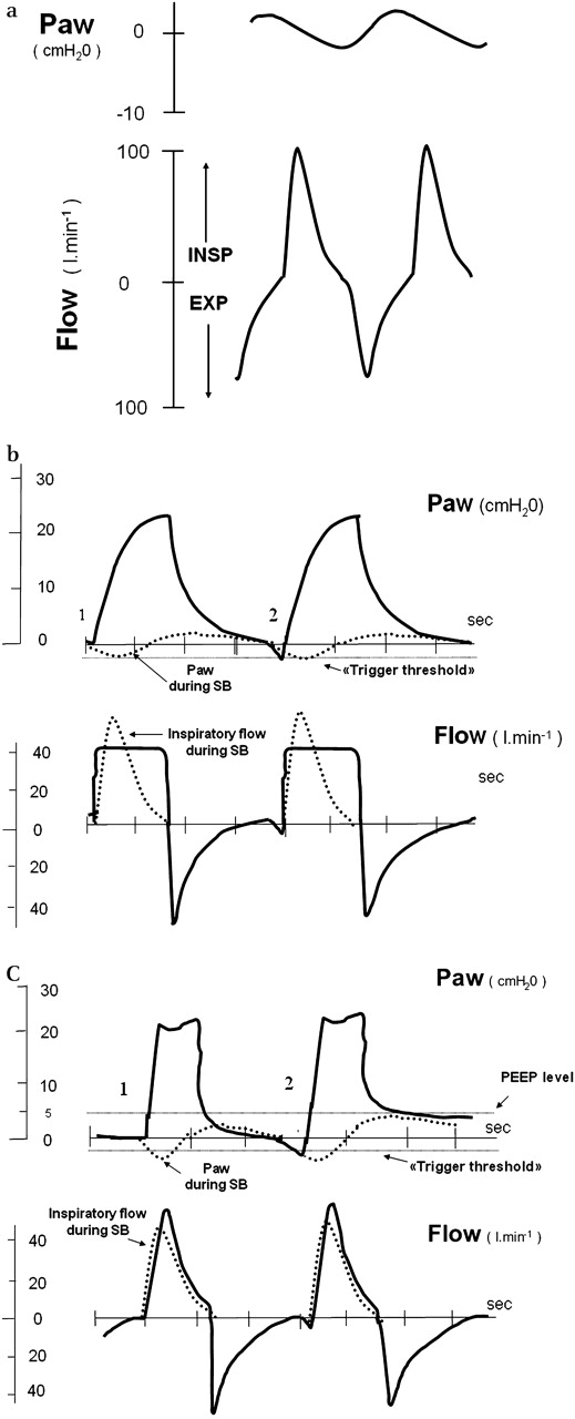
Flow and mask pressure during (A) spontaneous breathing, (B) volume-targeted ventilation, (C) pressure-targeted ventilation. 1, controlled cycle; 2, assisted cycle. In (B) and (C), dashed lines represent spontaneous breathing (SB) traces from (A). Paw, pressure in the airways; PEEP, positive end expiratory pressure.
Pressure-targeted ventilation (PTV)
In this modality the ventilator is set to deliver airflow by generating a predefined positive pressure in the airways for a given time. Airflow is therefore adjusted in order to establish and maintain a constant Paw. Constant analysis of the flow rate and airway pressure determines the flow variations necessary to maintain a flat or ‘square wave’ pressure. Flow is brisk at the beginning of inspiration when the gradient between the circuit pressure and the pressure target is large. As this gradient narrows the flow decelerates until driving pressure no longer exists and flow ceases (figure 2).
Thus, for a given patient, the volume delivered is not fixed and will depend on the interaction between the preset pressure, patient inspiratory effort, the physical characteristics of the respiratory system (C and R) and inspiratory time. An important advantage of PTV is the ability to compensate for mild to moderate leaks.
A comparative analysis of the two modalities and of corresponding flow and pressure patterns is summarised in table 1.
Comparison between pressure and volume-targeted ventilators
Volume-targeted pressure ventilation
A limitation of pressure ventilation is that it cannot guarantee a tidal volume delivered to the patient. Volume targeting is a feature available in some new ventilators that could allow this limitation to be overcome. This hybrid modality combines features of pressure and volume ventilation. The ventilator estimates the delivered tidal volume and adjusts its parameters to ensure a predetermined target tidal volume. Some ventilators adjust a target volume within each cycle (each breath starts as a pressure-limited breath and if the set tidal volume is not reached, the breath converts to a flow-cycled modality by prolonging the inspiratory time), but most of them progressively adjust the pressure level during several cycles to provide a tidal volume as close as possible to the target volume set by the clinician.
Whether this feature improves the effectiveness of ventilation is not yet clear.23 24 Different algorithms used to provide volume-targeted pressure are shown in figure 3.

Different algorithms of volume-targeted pressure. Pressure, volume and flow recording traces using (A) an algorithm that progressively adjusts the pressure support level over several cycles between a preset range of pressure to achieve a target tidal volume (Vt) and (B) an algorithm that adjust a target volume intracycle. Note that, in this case, each breath starts as a pressure-limited breath and if the set Vt is not reached, the breath converts to a flow-cycled mode by prolonging the inspiratory time. In both cases the ventilator is able to estimate the cycle to cycle Vt effectively delivered to the patient and to adjust the ventilatory cycle to ensure a target Vt. Press, inspiratory pressure. Reproduced with permission of A Cuvelier.
Recent ventilator modes: matching physiology and practice?
Conventional ventilation delivers fixed pressure or volumes rates. Some recent modes use a more physiological delivery of assistance following the spontaneous inspiratory signal coming from the patient. Hence, they are supposed to improve patient ventilator synchronism.
One of these modes, proportional assisted ventilation (PAV), relies on the inspiratory flow of the spontaneously breathing pattern and adds a fixed proportional assistance. If the patient's flow increases the assistance increases proportionally. The degree of assistance is determined by the practitioner based on estimations of the patient's resistance and compliance.25 Even if PAV proves to work according to theoretical assumptions, clinical studies using this mode both in invasive ventilation and NIV failed to show any clear advantages of this mode over others.26–29
The other mode, called neurally-adjusted ventilatory assist (NAVA), relies on the EMG of the diaphragm recorded from oesophageal electrodes. Ventilatory assistance is therefore not influenced by factors such as airway resistance, elastance, intrinsic positive end expiratory pressure (PEEP) or air leaks.30 This modality, available only in critical care ventilators, was tested only in patients under invasive ventilation. As there are very few data on NAVA application, its clinical impact cannot be assessed at present.
Influence of ventilator cycling: who controls the ventilator and who controls ventilation?
Appropriate settings are decisive to obtain optimal patient-ventilator synchrony.8 When delivering mechanical ventilation there are two ventilatory pumps acting together: the ventilator on the one hand and the patient's own respiratory muscles on the other. These two pumps may work in harmony but, in fact, they can interact in any number of ways, many of which will create problems rather than solving them. Hence, patient-ventilator asynchrony is quite common in patients during NIV.31 32 Asynchronies may occur at two levels: during inspiratory triggering in situations in which there is a mismatch between patient inspiratory effort and ventilator triggering (ie, ineffective inspiratory effort, double triggering or autotriggering); or during cycling from inspiration to expiration when ventilator cycling does not coincide with the end of patient effort (ie, premature or delayed cycling).32
Thus, the most logical approach to explain how a ventilator acts is to analyse the different phases of a typical positive pressure ventilatory cycle (figure 4).

The ventilatory cycle as seen from a mask pressure-time tracing. I to E, inspiratory to expiratory; PEEP, positive end expiratory pressure.
We will start this section by reviewing the triggering and cycling modes which refers, respectively, to how inspiration and expiration start and stop.
How is the onset of ventilator inspiration determined?
The patient can control the initiation (eg, triggering) and the end of inspiration (eg, cycling into expiration) or, on the contrary, neither of them, and the ventilator controls the initiation and the end of inspiration.
Spontaneous mode (S)
In this mode, only available in PTV, the patient controls the beginning and end of inspiration. Inspiration starts when the ventilator is triggered by the patient. The pressure is maintained as long as a minimal preset inspiratory flow is occurring. End of inspiratory assistance (eg, cycling from inspiration to expiration) occurs when inspiratory flow reaches a predetermined percentage of peak inspiratory flow. In this mode, a targeted inspiratory pressure, inspiratory trigger sensitivity and a percentage threshold of peak flow for cycling to expiration (see below) must be selected. In some ventilators these can all be set by the clinician whereas in others only the inspiratory pressure can be set. Because each cycle is terminated by a flow criterion rather than by volume or time, the patient retains control of cycle length as well as its depth and flow profile This mode is also called ‘pressure support ventilation’ (PSV) (figure 5A).
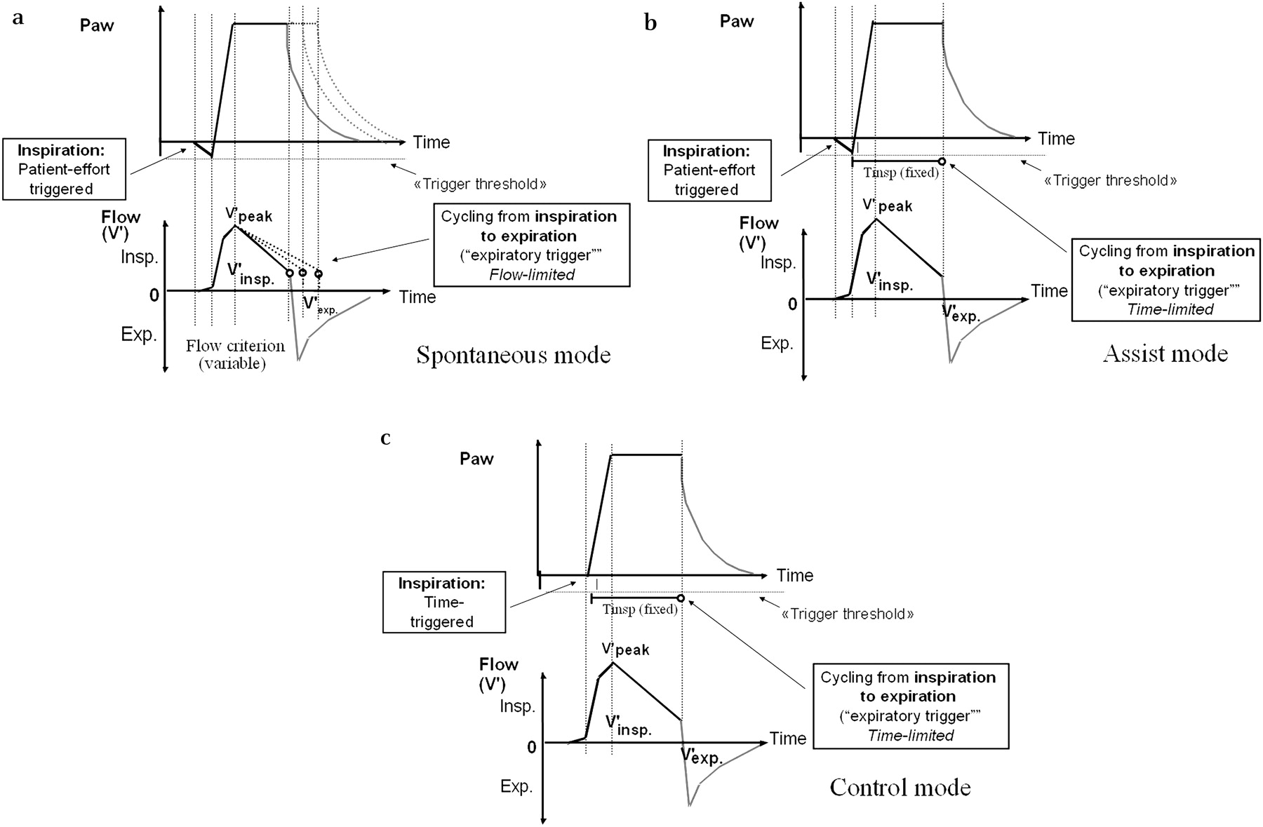
Different modes of inspiratory and expiratory triggering. (A) Spontaneous mode: the patient controls the beginning and end of inspiration. Inspiration starts when the ventilator is triggered by the patient and cycling from inspiration into expiration occurs when the inspiratory flow reaches a predetermined percentage of peak inspiratory flow. This mode is also called ‘pressure support’ and, in some ventilators, ‘spontaneous (S) mode’. (B) Assisted mode: the patient controls the onset of inspiration but the end of inspiration is time triggered. When a backup respiratory rate (RR) is preset, this mode is called assist/control. In this last mode, if the patient's RR is lower than the preset ventilator backup RR, the system moves to control mode. (C) Control mode: there is a preset automatic cycle based on time. The ventilator controls the beginning and end of inspiration and thus the RR. In some ventilators this mode is also called ‘timed (T) mode’. Paw, pressure in the airways.
Assist mode (A)
In this mode the patient controls the onset of inspiration but the inspiratory length is regulated by the operator. The clinician must select a targeted volume or pressure, an inspiratory:expiratory (I:E) ratio or an inspiratory time and an inspiratory trigger sensitivity (figure 5B).
Assist-control mode (A/C)
This mode operates as an assist one but also allows selecting a back-up respiratory rate (RR). If the patient's spontaneous frequency is lower than the preset ventilator back-up RR, the system moves to control mode. Therefore, this mode allows the patient to trigger the ventilator but also grants a minimum back-up rate. In this mode the clinician must select the same settings as in assist mode but also add a back-up rate.
Control mode (C)
In the control mode there is a preset automatic cycle based on time. The ventilator controls the beginning and end of inspiration and thus the RR. One therefore expects the ventilator to capture and inhibit the patient's respiratory centre and for the patient to follow the settings imposed by the ventilator. In this mode the clinician must select a targeted volume or pressure, a fixed RR and an I:E ratio or inspiratory and expiratory durations. With this mode, the entire work of breathing is supposed to be performed by the ventilator. In some ventilators this mode is also called ‘Timed (T) mode’ and is rarely used (figure 5C).
A particular combination of these modes is available in some NIV ventilators. This mode, called S/T, is basically a PSV that provides a back-up rate. In this particular mode, cycling from inspiration to expiration is flow-limited in patient-triggered cycles and switches to time-limited when the patient's spontaneous RR falls below the back-up RR, and also when the inspiratory time exceeds a predetermined maximal length during S cycles (see below). A patient-triggered cycle can be seen in curves of ventilation as a negative inspiratory deflection in pressure and flow curves (see trace 2 in figures 2B and C).
Type of trigger
As described above, in the A and A/C modes the ventilator has to recognise the patient's inspiratory effort. This is called triggering function. Classically, NIV devices have two types of triggers. The first, called ‘pressure-based’ trigger, present in old ventilators, is based on a drop in proximal airway pressure and requires a closed circuit. The second, called ‘flow-based’ trigger, present in almost all recent ventilators, is based on detection of an inspiratory flow in the presence of continuous flow washing out the circuit during expiration. Asynchrony during inspiratory triggering is quite common during sleep in patients during NIV and may compromise ventilatory efficacy and sleep quality. It is mainly influenced by the delay duration (that can vary between different ventilators33), the trigger sensitivity and the amount of inspiratory effort (which depends itself on respiratory drive and muscle strength).31
Ventilators that use flow triggering generally have the shorter trigger delays.34 35 Leaks may greatly affect trigger function, either by preventing the detection of patient inspiratory effort (leading to ineffective inspiratory effort) or by mimicking an ‘inspiratory flow’ (when using flow triggering) or dragging the EPAP level below the trigger threshold (when using pressure triggering), both situations being able to lead to autotriggering (figure 6).

Example of autotriggering. Autotriggering is defined as the occurrence of at least three consecutive pressurisations at a ventilator frequency of >40/min not synchronised with patient respiration.43
The newer technologies (microprocessors, servo valves and fast blowers) have substantially improved trigger responses. Moreover, adjustable inspiratory trigger is an option presently available in most home ventilators. Some of these also propose automated complex trigger algorithms (‘flow waveform method of triggering’) in which the flow-time waveform is used to trigger the ventilator. The respective advantages of these sophisticated trigger systems have not been assessed in rigorous studies. It must be emphasised that some adjustable trigger devices are graduated by arbitrary units which makes them difficult to use.
Pressurisation rate
As correct pressurisation is essential to decrease inspiratory effort and improve synchronisation, during this phase inspiratory flow should be sufficient to match inspiratory demand.36 Circumstances influencing the rate of pressurisation are the level of ventilatory support, the amount of time required to reach the pressure target (pressurisation slope, also called ‘rise time’), the compliance and resistance of the respiratory system and the patient inspiratory effort. Studies comparing different ventilators also emphasise the influence of the type of device on pressurisation, in particular in situations of high inspiratory demand.33
A faster rise time has been shown to unload respiratory muscles more completely.36 As the slope becomes flatter, the machine delivers lower flow rates and the patient's work of breathing increases.36 On the other hand, it must be emphasised that, if slow pressurisation could increase inspiratory work, an excessive peak flow could also have adverse effects as it may increase the sensation of dyspnoea,37 induce double triggering32 and lead to high peak mask pressure, favouring leaks.
Ability to sustain the inspiratory plateau
The inspiratory pressure level is one of the main determinants of the efficaciousness of NIV. Determination of the optimal level is the result of balancing two opposing aims: the desire to provide effective minute ventilation and the need to minimise leaks and discomfort caused by excessive inspiratory pressure. It must be emphasised that, even if recent ventilators have a good capability to compensate mild to moderate leaks, greater leaks may compromise the ability of the device to attain a desired level of inspiratory pressure.
Cycling from inspiration to expiration
Switching from inspiration to expiration can be time-cycled or flow-cycled. In time-cycled modes, ventilators use time criteria chosen by the clinician (figure 4B). In flow-cycled modes, cycling occurs as inspiratory flow decreases to a predetermined percentage of the peak inspiratory flow which is supposed to indicate the end of inspiratory effort (figure 4A).
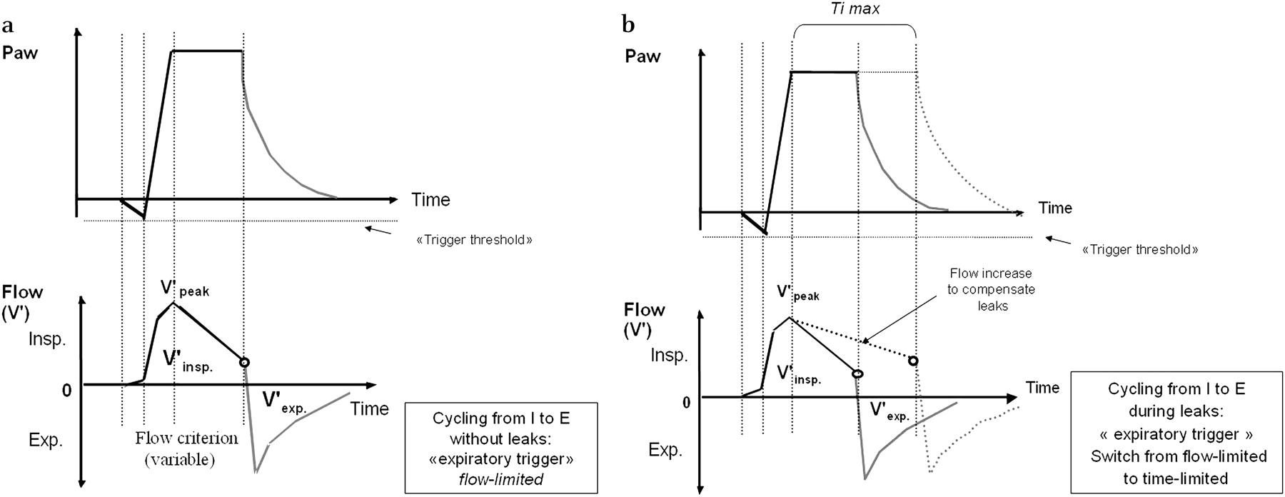
The criteria used to end inspiration may have a clinically relevant impact on the quality of ventilation. Ideally, cycling should coincide with the end of patient effort. However, synchronisation between the end of neural inspiration and ventilator cycling into expiration is mainly determined by respiratory mechanics, moving from a premature cycling in restrictive diseases to a late cycling in obstructive ones.33 38 Moreover, when flow cycling is used, leaks may also delay switching into expiration because, in an attempt to maintain pressure, the flow rate is maintained above the level at which cycling into expiration occurs (figure 7). Both these conditions may lead to patient-ventilator expiratory asynchrony.38
In older ventilators cycling into expiration was fixed at 25% of peak flow, but more recent ventilators offer adjustable values. This may allow tailoring settings to the patient's underlying condition. For instance, Tassaux et al demonstrated in patients with chronic obstructive pulmonary disease during invasive ventilation that increasing cycling from inspiration to expiration from 10% to 70% of peak flow (this means shortening inspiration to allow a greater expiratory time) was associated with a marked reduction in delayed cycling and intrinsic PEEP.39
Finally, additional mechanisms proposed by some ventilators control the end of inspiration to prevent undesirable prolongation of inspiratory time. Sudden increases in pressure (that can be assumed as secondary to an active expiratory effort) produce early cycling to expiration in almost all the devices. Another mechanism is to limit maximal inspiratory time. This maximal inspiratory time acts as a safety feature to prevent unsuitable lengthening of inspiratory duration and is highly variable in different ventilators. Moreover, it may be adjustable for some devices (figure 7).

Impact of leaks on inspiratory to expiratory (I to E) cycling during pressure support ventilation (S in bilevel devices) mode (A) without leaks and (B) with leaks. Note that during leaks the flow increases to compensate for them, prolonging the inspiratory time (dashed lines) and switching to time-limited if maximal inspiratory time (Timax) is available. Interestingly, Timax is adjustable in most recent non-invasive ventilation devices. Paw, pressure in the airways.
PEEP level
In patients with obstructive disease, intrinsic positive end expiratory pressure (iPEEP) can reduce the effective trigger threshold leading to a significant delay between the onset of patient inspiratory effort and ventilator triggering or even to ineffective efforts. In these cases, providing an external PEEP may counterbalance iPEEP, improving patient-ventilator synchrony. PEEP is an above atmospheric (positive) pressure applied during expiration. It is called EPAP in some ventilators. If PEEP is used, the pressure will never come back to zero. Indeed, IPAP results from pressure support plus PEEP. As a result and with regard to the ventilator category (ICU or home ventilator), the PEEP setting may interfere with either pressure support or IPAP levels. In fact, ICU ventilators propose PEEP and pressure support settings, but PEEP and IPAP settings are usually associated on home ventilators. Thus, the PEEP setting increases the IPAP level on ICU ventilators and decreases the pressure support level on home ventilators. Providing an external PEEP during NIV has many other theoretical advantages: flushing CO2 from the deadspace, preventing rebreathing within the mask, preserving the airway patency in patients with unstable UA during sleep and recruiting alveoli. Unnecessary increases in PEEP levels must be avoided because inspiratory pressure must be increased in parallel if inspiratory assistance is to be maintained, and this can lead to intolerance and favour leaks. Leaks, if important, may make it impossible to maintain the set EPAP level.
NIV modalities and settings: time to define a standardised nomenclature?
As underlined above, there is a wide variety of existing terminologies describing NIV modalities without a common nomenclature and with a lot of confusion. It even happens that the same acronyms correspond to different modalities and that identical modalities are called differently.
As ventilators can be categorised by the way that they deliver gas flow and by how they shift from inspiration to expiration and back, the answer to three basic questions may be used as a guide to simplify terminology:
What is the controlled variable (pressure or volume)?
What causes the start of ‘ventilator inspiration’? It can be either the patient (triggering) or the machine.
What determines the end of inspiration? It is either based on time (time-cycled) or determined by the patient (flow-cycled).40
Table 2 summarises the technical characteristics of NIV ventilators and their influence on monitoring tracings.
Summary of technical characteristics of NIV ventilators and their influence on traces semiology
Impact of ventilator mode, settings and function on monitoring during NIV
Using NIV, being connected to a ventilator is not synonymous to being ventilated. It is therefore essential to assess NIV efficacy. A first step is to perform a clinical assessment with the patient awake. However, as NIV is generally applied for several hours during sleep, more specific and sophisticated monitoring is needed to assess overnight patient-machine ‘agreement’ and efficacy of ventilation.
The most available and simple monitoring can be done from flow and pressure curves. Examination of these tracings can give useful information to evaluate whether the settings chosen by the operator are the right ones for that patient. However, as NIV allows a large range of ventilatory parameters and settings, it is mandatory to have accurate information about these issues to better understand the interplay between the patient and the ventilator. Ventilatory modality, mode of triggering, pressurisation slope, use or not of PEEP and type of exhalation as well as ventilator performances may all have semiological consequences on ventilatory traces. Leaks and variations in UA resistance may, in turn, modify these patterns.
Understanding the influence of non-invasive ventilator functions on the appearance of abnormal respiratory events and an appropriate mastery of the available tools for monitoring NIV during sleep are prerequisites to a better understanding of how patients and ventilators interact. These issues are analysed in two other articles in this review series.41 42
Acknowledgments
The authors thank ADEP Assistance (Paris, France) for the organisation of the workshops of the SomnoNIV Group.
References
Footnotes
Competing interests None.
Provenance and peer review Commissioned; externally peer reviewed.