Article Text

Statistics from Altmetric.com
Clinical presentation
A 46-year-old man was admitted with confusion and lower limb weakness that had developed over 2 weeks. A history of chronic productive cough was noted. Significantly, 4 years previously he had been investigated for cough and left apical lung consolidation. No evidence of Mycobacterium tuberculosis was found at the time and this was not investigated further. The only other relevant history was of heavy ethanol intake and self-neglect.
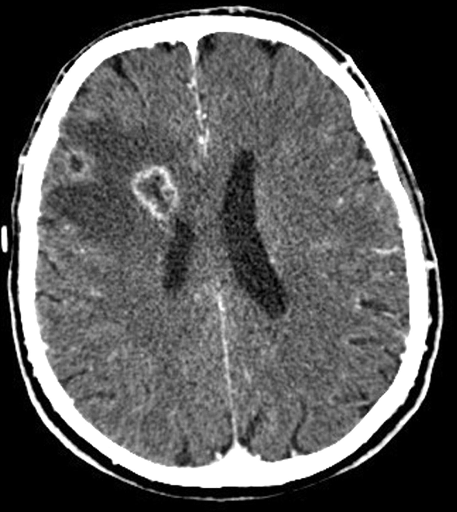
The patient was febrile (38.6°C), confused and unwell. He had evidence of finger clubbing, poor oral hygiene and signs of consolidation in the left lung. Early bilateral papilloedema and a flaccid paralysis in the lower limbs were noted. Admission investigations identified a raised white cell count of 21.5×10⁹/l (neutrophils 19.5). Multiple sputum cultures were negative. There was no reaction to a Mantoux (5 units PPD) test. Antibodies to HIV were not detected. A chest radiograph (fig 1) and CT scan showed left apical consolidation and volume loss. A CT scan of the brain (fig 2) showed two ring-enhancing lesions in the right frontoparietal lobe. An MRI scan revealed a ring-enhancing lesion in the lumbar spine.

Chest radiograph showing left apical consolidation, volume loss and pleural thickening.

CT scan of the brain showing ring-enhancing lesions in the right frontoparietal lobe with surrounding oedema and mass effect.
Craniotomy and excision of a cerebral lesion was performed, with concurrent bronchoscopy and bronchoalveolar lavage (BAL). The airways were inflamed with inspissated yellow secretions visible on the left. Histological examination of the cerebral biopsy specimen was consistent with a cerebral abscess, but no granulomatous inflammation was identified. All cultures from the BAL fluid and cerebral abscess were negative.
Question
What further diagnostic technique may aid in pathogen identification?
Answer
Broad-range bacterial PCR analysis was performed on the cerebral biopsy specimen1 and 16S rDNA of Prevotella spp was detected. A diagnosis of chronic Prevotella spp infection was made. Prevotella spp are a group of Gram-negative anaerobes that colonise normal human mucosae and are recognised causes of oral, head and neck, and respiratory infection. Signs of chronic suppurative infection are typically present. Deep-seated pure Prevotella infections—notably CNS abscesses—may occur, probably as a result of haematogenous spread.2 Infections ensue when host factors (such as malnutrition and self-neglect) disrupt the normal commensal relationship.2 Chronic poor oral hygiene, alcoholism and a consequent risk of inhalation therefore put our patient at high risk of anaerobic pleuropulmonary infection with ensuing haematogenous dissemination.
This case illustrates the potential role of molecular identification techniques in diagnosing unusual causes of respiratory infection. Despite culture of blood, sputum, bronchoalveolar lavage fluid and brain tissue, no pathogen was isolated. The difficulty in isolating and culturing anaerobic organisms is well recognised, with organism survival and recovery rate adversely affected by specimen handling.3 These infections are frequently polymicrobial and generally are diagnosed based on clinical presentation. RT-PCR is a rapid and highly sensitive diagnostic tool that has been used to identify a wider array of pathogens than is achieved with concomitant culture, and also enables identification of pathogens present in numbers below the lower detection limit of traditional culture.4 5
The patient was treated with metronidazole for 6 weeks and his respiratory symptoms and confusion resolved, although he has persistent lower limb paresis.
Acknowledgments
The authors thank Dr John Hartley, Great Ormond Street Hospital, London who performed the PCR analysis.
Footnotes
Competing interests None.
Patient consent Obtained.
Provenance and peer review Not commissioned; externally peer reviewed.
MGJ and SDM contributed equally to this paper.
Linked Articles
- Airwaves