Article Text

Statistics from Altmetric.com
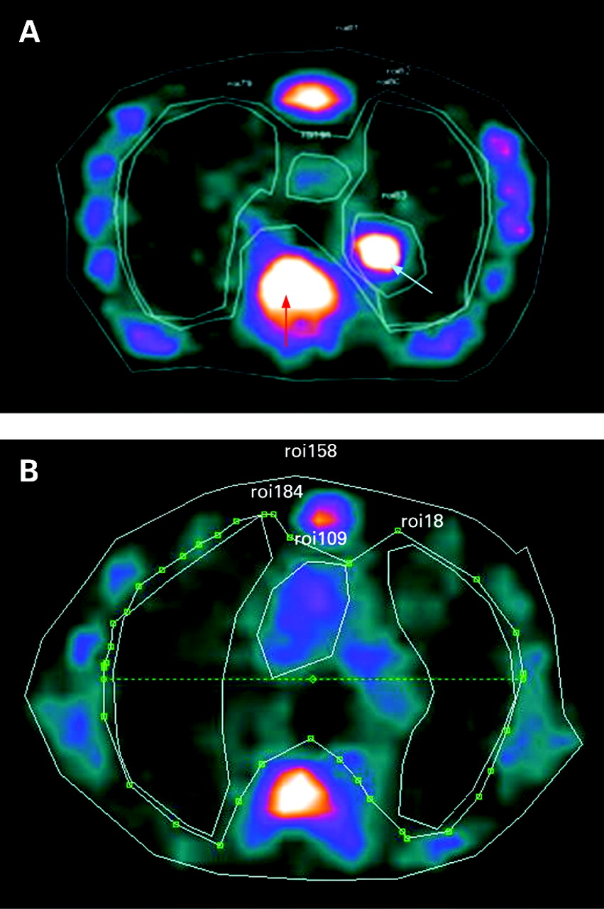
A 70-year-old man with severe smoking-related chronic obstructive pulmonary disease (COPD) was recruited to a research study to examine the trafficking of 99m-technetium (99mTc)-labelled neutrophils. These studies were undertaken in patients with stable moderate to severe COPD and designed to establish novel methodology (using autologous 99mTc-labelled neutrophils and single photon emission computed tomography (SPECT)) to quantify neutrophil trafficking and accumulation within the lungs of patients with COPD. At the time of scanning he was clinically stable with a C-reactive protein of 6 mg/ml and peripheral neutrophil count of 3.2×10⁹/l. He was a current smoker with a >100 pack-year history and had no other lung condition. His last COPD exacerbation was 4 months before the scan.
He was injected with 200 MBq 99mTc-labelled autologous neutrophils and underwent SPECT scanning at 45 min, 2 and 4 h post injection using a Picker Prism 2000 gamma camera. Following image acquisition, regions of interest were drawn around the cardiac, bone marrow and lung regions. As shown in fig 1A, the 4 h scan demonstrated unexpected and intense focal activity in the left lower lobe indicative of localised uptake of 99mTc-labelled neutrophils (a 4 h image from an age- and severity-matched current smoking subject with COPD is shown in fig 1B, demonstrating diffuse lung uptake only). Of note, at 4 h there is minimal activity present within the vascular compartment. Over the subsequent 48 h the patient developed symptoms of increased dyspnoea, cough, purulent sputum, fever and signs of focal consolidation confirmed on a chest radiograph. Moraxella catarrhalis was isolated from his sputum. The patient made a full clinical and radiological recovery following antibiotic administration.
Previous animal studies have shown that neutrophil migration into the lung following experimental infection is rapid in both onset and offset, with migration ceasing after 6 h.1 As a consequence, radiolabelled neutrophil imaging in patients with uncomplicated community acquired pneumonia is invariably negative.2 Focal activity can, however, be seen when neutrophil activity is monitored using 18F-labelled fluorodeoxyglucose (18FDG).3 We believe this is the first image of neutrophil uptake in a patient with pneumonia obtained using radiolabelled neutrophils and SPECT.
Learning point
Neutrophil migration into the lung in community acquired pneumonia is both rapid and transient such that white cell scanning using autologous 99mTc- or 111In-labelled neutrophils is almost always negative. Focal neutrophil activation, however, persists and can be detected using 18FDG-PET.
Linked Articles
- Airwaves