Article Text

Statistics from Altmetric.com
A 68-year-old woman was referred for investigation of pulmonary artery hypertension. Right heart catheterisation was performed and the tip of the catheter was positioned in the right middle lobe pulmonary artery under fluoroscopic guidance. A Swan–Ganz balloon was inflated to record a pressure of 78/23 mm Hg (mean 42 mm Hg). Immediately after the balloon was inflated, the patient experienced small volume haemoptysis.
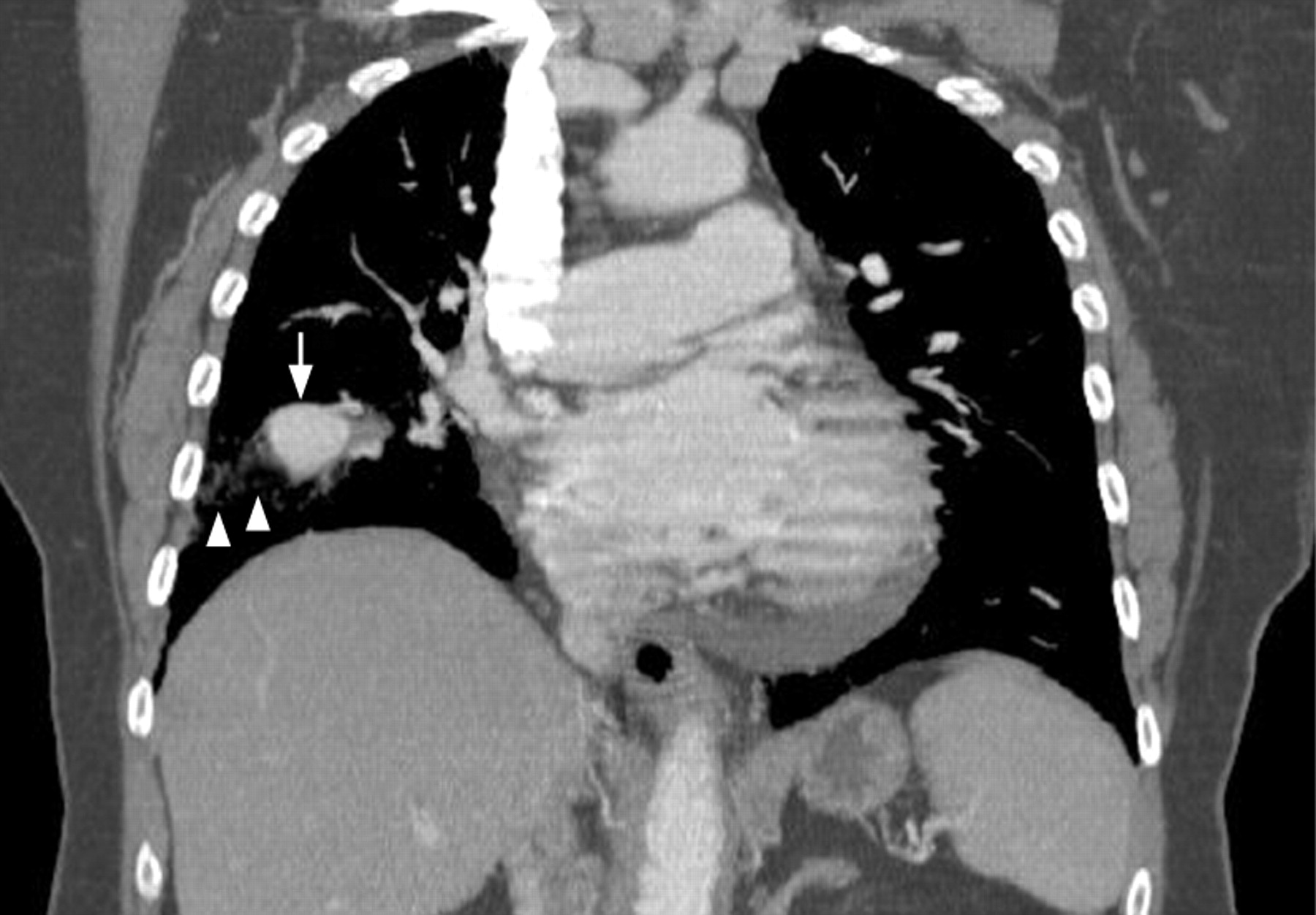
A CT pulmonary angiogram was performed later the same day to investigate the cause of her pulmonary artery hypertension. This showed a 3 cm pseudo-aneurysm arising from a right middle lobe subsegmental pulmonary artery with associated pulmonary haemorrhage (fig 1). The patient remained haemo-dynamically stable and was scheduled for elective embolisation of the pseudo-aneurysm. Before this, cardiac MRI was performed (6 days after catheterisation) to assess right ventricular dimensions, allowing the opportunity to visualise the pseudoaneurysm on the MRI scan (fig 2). A catheter angiogram was performed the following day with embolisation of the inflow artery by uncoated platinum coils (fig 3).


Learning points
The risk of pulmonary artery rupture with haemorrhage following Swan–Ganz catheterisation is 0.4% with pulmonary artery hypertension conferring increased risk.1
Most ruptures occur in the middle or lower lobe branches of the right pulmonary artery and carry a mortality rate up to 50%. Transcatheter embolisation is the treatment of choice.2
Footnotes
Funding: None.
Competing interests: None.
Patient consent: Obtained.
Linked Articles
- Airwaves