Article Text

Statistics from Altmetric.com
A 62-year-old male smoker was reviewed for increasing dyspnoea, hoarseness and stridor. The patient underwent a bronchoscopic examination of the airway that revealed two large ‘kissing’ fleshy tracheal lesions just below the vocal cords (figure 1). A biopsy was taken. The histological image (figure 2) was stained using a Congo Red Staining Kit which selectively demonstrates amyloid in the biopsy sample as bright pink and is marked ‘A’ in the image. The darker coloured cells represent airway epithelium. Pulmonary amyloidosis is rare and patients may present with tracheobronchial infiltration, parenchymal infiltration (amyloidoma), persistent pleural effusions or pulmonary hypertension. Symptoms of tracheobronchial amyloidosis include hoarseness, stridor, dyspnoea and overt airway obstruction. Invasive bronchoscopic therapies such as argon photocoagulation, bronchoscopic Nd:YAG laser debulking or surgical debulking may be required to relieve the obstruction.1
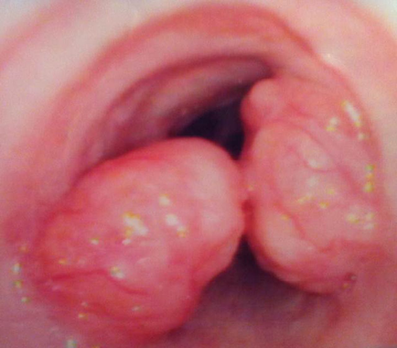
Two ‘kissing’ fleshy tracheal lesions.

Congo Red stained biopsy.
Footnotes
-
Competing interests None.
-
Provenance and peer review Not commissioned; externally peer reviewed.
Linked Articles
- Airwaves