Article Text

Statistics from Altmetric.com
An 18-year-old man presented to the emergency department of a community hospital with a cough, fever, night sweats and significant weight loss. He recently completed an antibiotic course for a lower respiratory-tract infection. Full blood count revealed a normal total white cell count with eosinophil count of 0.85×10⁹/L. C-reactive protein (CRP) was normal but erythrocyte sedimentation rate (ESR) was 100 mm/hour. Chest radiograph demonstrated bilateral perihilar reticulonodular opacification with peripheral sparing (figure 1A). Immunoglobulin E was mildly elevated at 175UI/mL; other immunoglobulin levels, viral and autoantibody panels were normal. High-resolution CT thorax revealed diffuse centrilobular and peribronchovascular ground-glass opacification throughout both lungs with subpleural sparing (figure 1B/C). On further questioning, the patient disclosed a history of extensive vaping in the preceding 6 months including nicotine and tetrahydrocannabinol-containing products. He was referred to a specialist interstitial lung disease clinic where pulmonary function tests demonstrated normal spirometry but a reduced diffusing capacity of the lungs for carbon monoxide (DLCO) of 60.3% predicted. A bronchoscopy was arranged 4 weeks after his initial presentation, and a repeat chest radiograph was performed immediately prior to the procedure demonstrating marked resolution in parenchymal abnormalities (figure 2A). Due to the improved radiological changes, no transbronchial biopsies were performed, but bronchoalveolar lavage (BAL) revealed a cell differential of 66% macrophages, 27% lymphocytes, 4% neutrophils and 3% eosinophils (figure 2B). Approximately 25% of alveolar macrophages were foamy lipid-laden, demonstrated by neutral lipid containing droplets evident on Oil-Red-O staining (figure 2C). Though the cytological appearances varied slightly from recent reports of acute presentation, the resolution on chest radiograph and presence of a significant proportion of lipid-laden macrophages were considered to represent a resolving vaping associated lung injury. Following cessation of all vaping products, the patient improved and DLCO had markedly improved 8 weeks after initial presentation.
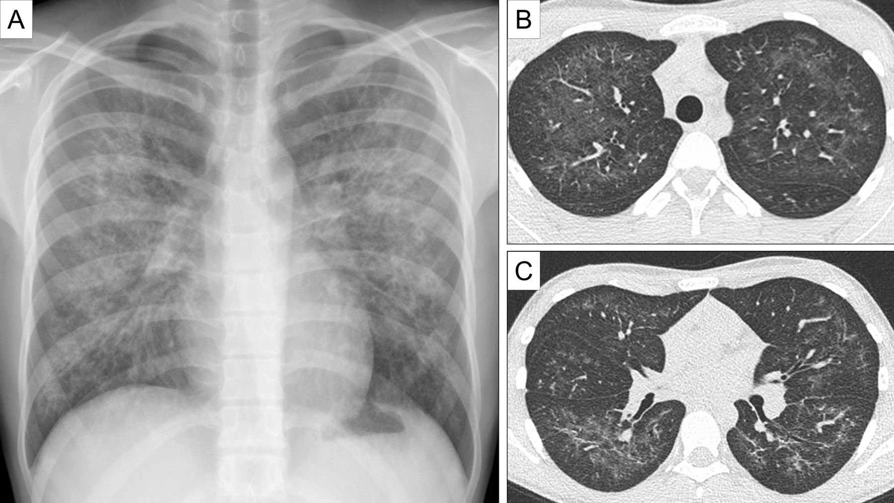
(A) Chest radiograph demonstrating extensive bilateral perihilar reticulonodular opacification with peripheral sparing. (B,C) Axial chest CT images demonstrate extensive bilateral centrilobular and peribronchial ground glass opacification with subpleural sparing, slightly more confluent in the lower zones.

(A) Chest radiograph demonstrating complete interval resolution of the perihilar reticulonodular opacities. (B) Bronchoalveolar lavage (BAL) cytology stained with MGG ×400 magnification demonstrating foamy macrophages, some multinucleated (yellow arrow). (C) Alveolar macrophages stained in red with ORO ×400 magnification demonstrating presence of excess neutral lipid in alveolar macrophages. MGG, May Grunwald Giemsa stain; ORO, Oil-Red-O stain.
The presence of airway lipid-laden macrophages can indicate aspiration of exogenous lipoid material or be present in pulmonary alveolar proteinosis syndrome. However, in cases of heavy use of electronic cigarettes, this may be a useful marker of the recently described syndrome: electronic cigarette and vaping associated lung injury (EVALI).1 Though the pathophysiological mechanisms are unclear, the presence of lipid-laden macrophages has been reported in many EVALI cases. Cases have similarly demonstrated peripheral blood eosinophilia (ranging from normal levels to 2.9×10⁹/L) elevated ESR (ranging from 60 to 128 mm/hour) and elevated levels of lipid laden macrophages on BAL assessment (ranging from 25% to 75%).1 Vitamin E acetate is the synthetic ester of tocopherol and acetate and is widely used as a thickening agent in vaping products. It is proposed that when inhaled, vitamin E acetate may interfere with the ability of pulmonary surfactant to maintain surface tension in alveoli, and when heated, it may form ketene, a known lung irritant.2 Recent animal model studies have demonstrated elevated levels of BAL lipid-laden macrophages on Oil-Red-O stain in mice exposed to aerosols generated from vitamin E acetate.3 This case highlights the utility of BAL and Oil-Red-O staining in the evaluation of EVALI and how it may be helpful even during resolution of imaging findings.
Footnotes
OO and KS are joint first authors.
Twitter @keshavs21
Contributors All authors contributed to the conception, data collection and writing of the manuscript.
Funding The authors have not declared a specific grant for this research from any funding agency in the public, commercial or not-for-profit sectors.
Competing interests None declared.
Patient consent for publication Obtained.
Provenance and peer review Not commissioned; externally peer reviewed.
Data availability statement Data are available upon request.