Article Text

Statistics from Altmetric.com
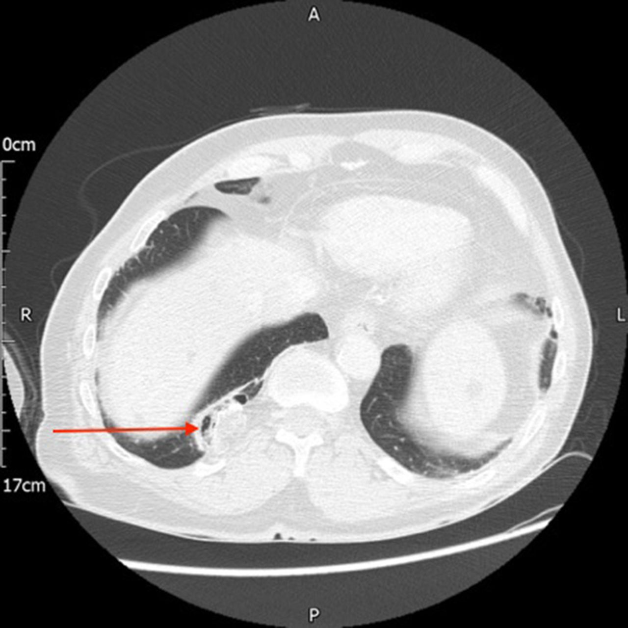
A 75-year-old man, with hereditary multiple exostoses, had a positron emission tomography (PET)-CT scan for follow-up of an excised chondrosarcoma. Incidentally, he was found to have widespread subpleural cystic change (figure 1A and B) and a cystic right lower lobe lesion (figure 2) adjacent to a rib exostosis. He was referred to the urgent respiratory clinic on the basis of these unusual subpleural cystic findings and concern that the cystic right lower lobe lesion might represent a lung adenocarcinoma. He had a significant smoking history (40 pack-years). Follow-up scans have shown gradual size increase of the subpleural cysts and a PET-CT scan showed the lesion to be fluorodeoxyglucose-negative and likely to represent a focal area of atelectasis or fibrosis associated with the adjacent rib osteochondroma.
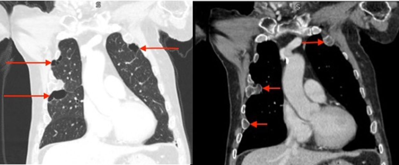
Coronal imaging showing cystic change on lung windows (A) and exostoses on soft tissue windows (B).

Axial image of right lower lobe lesion associated with rib exostosis on lung windows.
Discussion
Osteochondroma is the most common benign tumour of the bone, with costal manifestations in 1%–2% accounting for 50% of benign rib tumours and 8% of rib tumours overall. Costal osteochondroma usually affect children and young adults and can be idiopathic (solitary costal exostosis) or be part of a syndrome (hereditary multiple exostoses).
The presence of costal exostoses has implications for the intrathoracic viscera—they have been associated with numerous case reports of pneumothorax,1 haemothorax,2 3 pleural effusion, diaphragmatic laceration and pericardial and pleural thickening. Haemothorax may result from direct vascular trauma caused by the sharp tip of the exostosis or by spontaneous rupture of dilated vessels in the pleura arising from chronic inflammation caused by constant friction of the exostosis on the pleural surface during respiration. The subpleural cystic changes seen in our case have never been previously described, but may form part of the spectrum of a chronic inflammatory or traumatic process and may contribute to the pathogenesis of pneumothoraces and haemothoraces.
Footnotes
Contributors Manuscript prepared and written by MT and DA. Manuscript preparation overseen and edited by CB, AM and FG.
Competing interests None declared.
Patient consent Obtained.
Provenance and peer review Not commissioned; externally peer reviewed.
Linked Articles
- Airwaves