Article Text

Statistics from Altmetric.com
We have read with great interest the comments by Dr Persson1 on our recent paper in Thorax,2 in which we showed that clinical control of asthma associated significantly with lower numbers of activated eosinophils in the bronchial wall, yet only weakly with sputum eosinophils. As the number of eosinophils in biopsies did not associate with clinical control of asthma, we speculated that activation of eosinophils (measured as eosinophil protein X (EXP)-immunopositive pixels per area) in bronchial biopsies reflects the level of disease control better than the number of eosinophils itself.2 As lysis of activated eosinophils and degranulation of toxic eosinophil proteins may damage the surrounding tissue,3 Persson wondered whether EPX immunopositivity in our biopsies associated with epithelial fragility, particularly in uncontrolled asthma.
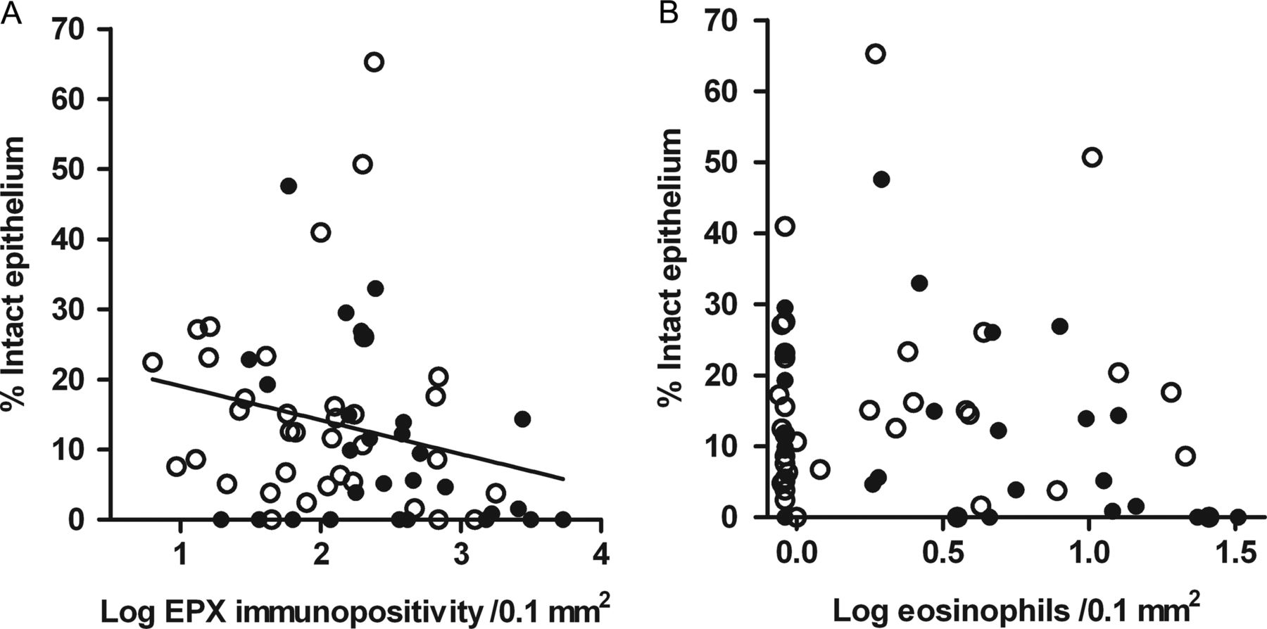
In line with Persson's hypothesis, the percentage of intact epithelium correlated negatively with EPX immunopositivity (Spearman's r=−0.30, p=0.016), whereas there was no significant correlation with the number of eosinophils in bronchial biopsies (Spearman's r=−0.12, p=0.35) (figure 1). This was not due to effects of current smoking, which is associated with increased epithelial cell proliferation, goblet cell hyperplasia, as well as with reduced eosinophil numbers in bronchial biopsies in asthma,4 since we excluded current smokers from our analysis. An additional regression model adjusted for inhaled corticosteroid use and atopy confirmed that loss of epithelial integrity and higher EPX immunopositivity are significantly associated with uncontrolled asthma, yet not with numbers of airway wall eosinophils (data not shown).
Correlation between percentage of intact epithelium and eosinophil activation assessed by EPX immunopositivity (panel A) and number of eosinophils (panel B) in bronchial biopsies from non-smoking asthmatics. Open dots: controlled asthma (n=36), solid dots: uncontrolled asthma (n=29). Significant correlation between EPX immunopositivity and % intact epithelium in panel A (Spearman's r=−0.30, p=0.016). Subjects with uncontrolled asthma have less intact epithelium and more EPX immunopositivity (panel A).
Another question from Persson's letter was whether free granules locate in close proximity of denuded epithelium. Unfortunately, this ‘geographical’ relationship is very difficult to quantify in a reliable way. Moreover, we believe this specific question could be better investigated prospectively using an allergen provocation model; collecting blood, biopsies and sputum at regular time points; similar to what has been done in the past by Aalbers et al.5 In our existing dataset, the dynamics of transepithelial migration of eosinophils6 (tissue-lumen correlations) cannot be investigated in a reliable way.
In conclusion, our statistical analysis supports Persson's hypothesis that ongoing lysis of activated eosinophils contributes to uncontrolled asthma. Our previous publication and our current analyses support the notion that loss of epithelial integrity may serve an important role in this respect, since it is independently associated with loss of asthma control.
Footnotes
-
Contributors FF and NtH performed the analyses and wrote the answer. HKR, DSP, WT and MNH gave comments and revised the letter. All authors reviewed the final version.
-
Funding None.
-
Competing interests None.
-
Provenance and peer review Not commissioned; internally peer reviewed.