Article Text

Statistics from Altmetric.com
Ameneh Khatami and Emilie Huguon
A previously well adolescent from the tropical South Pacific island of Futuna was transferred due to a 3-month to 4-month history of intermittent fevers, anorexia, weight loss, lethargy and haemoptysis. A Mantoux test was negative. CT scan demonstrated a large mediastinal mass and lymphadenopathy with broncho-vascular compression, and bilateral pleural and pericardial effusions, (figure 1A). At admission, he was persistently febrile with non-tender cervical lymphadenopathy and hepatomegaly, and had moderate respiratory distress. The presumptive diagnosis was lymphoma.

Radiology investigations of patient with disseminated conidiobolomycosis. (A) CT scan on admission performed in prone due to respiratory distress demonstrating amorphous soft tissue mass filling the mediastinum and aortopulmonary window with compression of the right main bronchus (dotted arrow) and obstruction of the superior vena cava leading to accessory venous pathways filled with high-density contrast (solid arrow), bilateral pleural thickening and fluid (greater on the right) and irregular opacities in the right lung probably representing atelectasis. (B) CT scan after 3 weeks of antifungal treatment demonstrating several small, hypodense lesions throughout the liver (black arrows), multiple large wedge-shaped hypodense areas representing areas of infarction in the spleen and both kidneys and bilateral pleural effusions (greater on the left). (C) Positron emission tomography (PET) scan after 5 weeks highdose intravenous liposomal amphotericin demonstrating multiple foci of increased metabolic activity in the mediastinum, abdomen and soft tissues including in the right hand, bone involvement in the left 7th rib, a large left pleural effusion and non-uniform uptake in the cardiac muscle. (D) Follow-up PET scan after further 6 weeks intravenous liposomal amphotericin with addition of voriconazole and oral terbinafine, and 3 weeks of intravenous and oral prednisolone demonstrating significant clearance of infective foci.
Initial blood tests demonstrated haemoglobin 92 g/L; white cell count 16×109/L (neutrophils 11×109/L, lymphocytes 1.3×10⁹/L, eosinophils 1.2×10⁹/L); erythrocyte sedimentation rate 128 mm/h; C-reactive protein 313 mg/L; normal liver and renal function tests. After 1 week, he developed acute renal impairment with peak serum creatinine 368 µmol/L. Renal ultrasound scan (USS) demonstrated bilateral increased echogenicity. Cultures and cytology of the pleural effusion were non-diagnostic. Cervical lymph node biopsies demonstrated reactive histopathological changes and fibrosis, without evidence of malignancy.
David Isaacs
At this stage, the diagnosis is uncertain. The subacute presentation of a large mediastinal mass associated with pleural effusion, lymphadenopathy, fever and weight loss in a previously healthy adolescent is highly suggestive of malignancy, in particular, lymphoma. Mediastinal TB is an alternative differential diagnosis given the patient's background. The eosinophilia raises the possibility of fungal or parasitic infection, and in a patient from the tropics, Löffler’s syndrome associated with Strongyloides infection should be considered. Further information is required including a review of the history and deeper biopsy samples. Additionally, further tests for TB are necessary before this diagnosis can be confidently excluded.
Ameneh Khatami and Amanda Charlton
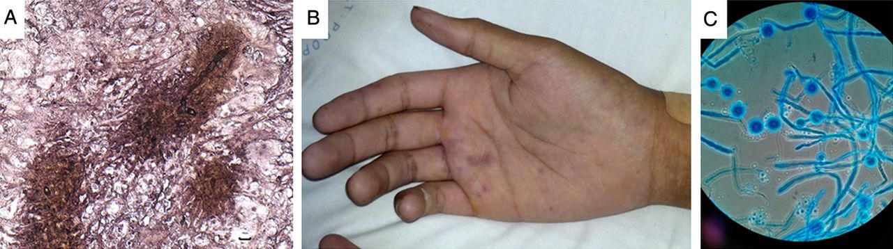
Detailed history revealed that symptoms started 2 weeks after the patient had sniffed a dead rat as part of a dare. Multiple sputum samples were negative for bacterial, fungal and mycobacterial culture, and there was no response to 2 weeks of broad-spectrum intravenous antibiotics. An interferon gamma release assay (IGRA) on whole blood was positive. Percutaneous core biopsies of the mediastinal mass demonstrated fungal hyphae in a fibroinflammatory background and Splendore–Hoeppli phenomena (SHP) (figure 2A).

Clinical, microbiology and histopathology images of patient with disseminated conidiobolomycosis. (A) Core biopsy of mediastinal mass showing branching fungal hyphae in the centre of Splendore–Hoeppli phenomenon. Methenamine sliver stain, bar is 10 µm. A short segment of hollow, fungal hypha (diameter 7.5 µm) is seen at the centre. (B) Tender violaceous lesions on the patient's right hand raised the possibility of endovascular infection. (C) Lactophenol Cotton Blue preparation of Conidiobolus incongruus cultured from a liver biopsy demonstrating spherical conidia with papillae.
Alison M Kesson and David Isaacs
A positive IGRA in an adolescent from a TB-endemic region is not unexpected. The histopathology suggests an invasive fungal infection, and the patient's raised eosinophil count would be consistent with this. The mediastinal mass is presumed to be a combination of mediastinitis and lymphadenopathy. The route of infection is presumed to be inhalation, however, it is unclear if the dead rat was the source of infection in this patient. There are no specific pathogenic fungi associated with decaying animal corpses. Imaging is necessary to look for involvement of the upper airways, and to exclude other sites of dissemination. Further investigation is required to exclude a primary immunodeficiency. Broad-spectrum antifungal therapy should be initiated, although care should be taken with intravenous liposomal amphotericin, given the patient's renal impairment.
Ameneh Khatami
Tests for chronic granulomatous disease and lymphocyte subset distributions were normal. HIV serology was negative. Serum immunoglobulin (Ig) E and IgG levels were elevated (10 800 IU/mL and 38 g/L, respectively). IgA and IgM levels were normal. Intravenous liposomal amphotericin was commenced on day 14 of admission at 3 mg/kg/day and increased to 9 mg/kg/day as renal function improved. Follow-up CT scan after 10 days antifungal treatment demonstrated some regression of the mediastinal mass; however, despite overall improvement in right lung aeration, there were new areas of lung consolidation and cavitation. No sinus or intracranial abnormality was detected. Eosinophilia peaked at 7×10⁹/L on day 35. As there had been a strong suspicion of malignancy initially, biopsy samples had not been provided for bacterial, fungal or mycobacterial cultures. The fungus was also not able to be identified using pan-fungal PCR on deparaffinised tissue from the mediastinal mass. The patient's respiratory status improved, however, he developed lymphoedema of the right arm, a new left-sided pleural effusion (demonstrated to be chyle when drained) and vasculitic lesions on his right hand (figure 2B).
Melanie Wong and Philip N Britton
The significantly raised IgE level is consistent with an invasive fungal infection, and the high serum IgG is consistent with immune activation. The paradoxical improvement in renal function following commencement of liposomal amphotericin may indicate direct fungal infection of the kidneys. Furthermore, the vasculitic lesions in the patient's hand raise the concern of endovascular infection. Scedosporium species are not covered by liposomal amphotericin and should be considered in cases of presumed angio-invasive fungal infection. Alternatively, these new lesions may be caused by a secondary bacterial infection or inflammatory vasculitis in the context of a profound systemic inflammatory response. Finally, all histopathology samples demonstrated extensive fibrosis which could have disrupted lymphatic drainage, causing the lymphoedema and chylothorax.
Ameneh Khatami
A transoesophageal echocardiogram showed no evidence of endocarditis or fungal invasion of the great vessels. Intravenous voriconazole was added, and intravenous antibiotics recommenced until multiple blood cultures returned negative. During the second month of admission, the patient developed severe epigastric pain, nausea and vomiting with ongoing high fever. Total parenteral nutrition (TPN) was started after 10 kg weight loss since admission. The dose of liposomal amphotericin was reduced to 6 mg/kg due to refractory hypokalaemia. Abdominal CT scan demonstrated numerous small, hypoechoic hepatic lesions (largest 12×7×5 mm) and large hypodense defects in the spleen and kidneys (figure 1B).
David Isaacs and Melanie Wong
The abdominal lesions noted on CT scan may be areas of infarction due to septic emboli or may be areas of abscess formation. Six weeks of high-dose liposomal amphotericin reduced the mediastinal mass and lymphadenopathy, without significant improvement in inflammatory markers, elevated IgE, eosinophilia or fever. Furthermore, inflammatory histopathological changes are disproportionate to scant fungal hyphae seen. It is likely that overwhelming systemic inflammation is contributing to the patient's ongoing symptoms. Adjunctive corticosteroids are effective in limiting organ damage resulting from intense inflammation associated with some pathogens, for example, tuberculous or Haemophilus influenzae type b meningitis or Pneumocystis jirovecii pneumonia,1 and should be considered for this patient. Corticosteroids have also been used in patients with granulomatous mediastinitis caused by Histoplasma capsulatum, where active inflammation of mediastinal lymph nodes can cause airway obstruction2; as well as an adjunct to antifungal therapy in treating refractory infections with invasive filamentous fungi and Streptomyces species in patients with chronic granulomatous disease.3
Ameneh Khatami and Amanda Charlton
Pulsed intravenous methylprednisolone was started on day 44 of admission, 2 mg/kg intravenous daily weaning over 3 weeks. This was associated with rapid defervescence, improved appetite and weight gain. Positron emission tomography (PET) scan revealed multiple foci of increased metabolic activity in the mediastinum, abdomen and soft tissues (figure 1C). Percutaneous biopsy of a liver lesion demonstrated damaged hepatocytes, fibrinous replacement and neutrophilic infiltrate with scant fungal hyphae. Conidiobolus incongruus was cultured from the liver biopsy (figure 2C), identified by sequencing the D1/D2 regions of the 28S ribosome subunit which demonstrated 98% homology to strain AF113547.
Alex C Outhred
Conidiobolus species are fungi of the order Entomophthorales, closely related to Basidiobolus spp.4 Human Conidiobolus infection was first reported in 1965.5 Almost all reported cases are from tropical or subtropical regions. Fungal inoculation occurs from minimal skin or mucosal trauma. There is no known association between contact with decaying animal corpses and Conidiobolus infection in humans. There is no consensus on optimal treatment. In vitro testing suggests Conidiobolus spp are highly resistant to most antifungal agents6 ,7; however, susceptibility testing has not been validated.
Alex C Outhred and David J E Lord
In vitro susceptibility testing of our Conidiobolus isolate at 3 laboratories gave discrepant results. The pattern reported by one was consistent with previous reports and suggested probable susceptibility to terbinafine, intermediate susceptibility to amphotericin and resistance to voriconazole (minimum inhibitory concentrations: amphotericin B=2 mg/L, anidulafungin=2 mg/L, micafungin >8 mg/L, caspofungin >8 mg/L, 5-fluorocytosine=32 mg/L, posaconazole=2 mg/L, voriconazole >8 mg/L, itraconazole >16 mg/L, fluconazole >256 mg/L, zone of inhibition around a terbinafine disc 30–40 mm). Therapeutic serum voriconazole levels were not achieved with up to 800 mg/day intravenous voriconazole. Treatment was switched to oral voriconazole, with omeprazole used to ‘boost’ serum levels and oral terbinafine was added.
Vomiting, abdominal pain and weight loss recurred as steroids and TPN were weaned. Angiography, performed because of two new focal liver lesions on USS, showed them to be aneurysms in the posterior right and left hepatic arteries and also demonstrated aneurysms in the gastro-duodenal and right hepatic arteries, although none outside the coeliac trunk. The cerebral vessels and coronary arteries were unaffected.
David Isaacs and Philip N Britton
The vascular complications in this patient are striking and include: arterial aneurysms, splenic and renal infarcts and peripheral vasculitic lesions. These may have resulted from fungal invasion of vessels with secondary septic emboli. Alternatively, they may have been due to an inflammatory vasculitis. The initial rapid renal impairment associated with parenchymal changes on renal USS and improvement in renal function with initiation of antifungal treatment suggest some direct fungal invasion.
Ameneh Khatami and David J E Lord
The three largest aneurysms were embolised, following which the patient's gastrointestinal symptoms slowly resolved. Persistent and marked hypertension required regular antihypertensives. A 6-week follow-up PET scan demonstrated dramatic clearance of hypermetabolic foci (figure 1D). Intravenous liposomal amphotericin was ceased after 13 weeks, when therapeutic levels of voriconazole were achieved. After 4 months inpatient treatment, the patient was discharged on oral terbinafine, voriconazole and amlodipine for a further 3 months. Two months following completion of treatment, he was well, asymptomatic and normotensive.
David Isaacs, Amanda Charlton and Melanie Wong
Entomophthorales are saprophytes found in tropical rain forests, soil, rotting vegetation and the gastrointestinal tract of insectivorous animals.8 Entomophthoromycota typically cause chronic cutaneous and subcutaneous granulomatous infections in immunocompetent individuals, whereas disseminated, angio-invasive Zygomycetes and Mucorales infections typically occur in immunocompromised hosts.9 Conidiobolus coronatus typically presents as a slowly progressive and disfiguring rhinofacial disease.9 Systemic conidiobolomycosis has previously been reported in one child and five adults.9 Disseminated disease is associated with non-coronatus species and progresses rapidly in contrast with rhinofacial disease. In these previous reports, infection was limited to the lower respiratory tract, neck and mediastinum. Four of the adults had comorbidities (immunosuppression in 3; 1 intravenous drug user). All adult patients died. A 15-month-old child with a posterior mediastinal mass survived following surgical excision and amphotericin B treatment.
Entomophthoramycosis induces an extensive eosinophilic and granulomatous reaction with variable neutrophilic infiltrates. SHP is the formation of radiate eosinophilic material, comprising antigen-antibody complexes, tissue debris and fibrin.10 It may prevent phagocytosis, leading to chronic infection. Alternatively, it may represent an effective host humoral response that limits fungal invasion, since absence of SHP has been associated with fatal angioinvasion in conidiobolomycosis.9 We saw SHP with clinical evidence of angioinvasion, suggesting that our patient's immune response did not prevent dissemination, perhaps because of a large initial inoculum. Nevertheless, subsequent aggressive antifungal therapy, in conjunction with a competent immune system, was able to control the infection. Our patient with was treated successfully for 6 months with a combination of agents.
Acknowledgments
We would like to thank Dr Catriona Halliday and Sue Sleiman at Centre for Infectious Diseases & Microbiology Laboratory Services, Institute for Clinical Pathology and Medical Research, Pathology West, Sydney; Kerry Weeks at Pacific Laboratory Medicine Services, Pathology North, Sydney; and Dr Sarah Kidd at National Mycology Reference Centre, South Australia Pathology, Adelaide for their help in identifying the Conidiobiolus incongruous isolate and for in vitro susceptibility testing. PNB acknowledges funding from Sydney Medical School and the Arkhadia Fund as Dean’s Fellow/Norah Theresa-Hayes Ratcliffe Lecturer.
Footnotes
-
Contributors All contributing authors were involved in the care of the patient. AK planned and prepared the first draft of the manuscript. All authors contributed to the development of the final version of the manuscript. AK is responsible for the overall content of the manuscript as guarantor.
-
Competing interests None.
-
Patient consent Obtained.
-
Ethics approval Sydney Children's Hospitals Network Human Research Ethics Committee.
-
Provenance and peer review Not commissioned; externally peer reviewed.
Linked Articles
- Airwaves