Article Text

Statistics from Altmetric.com
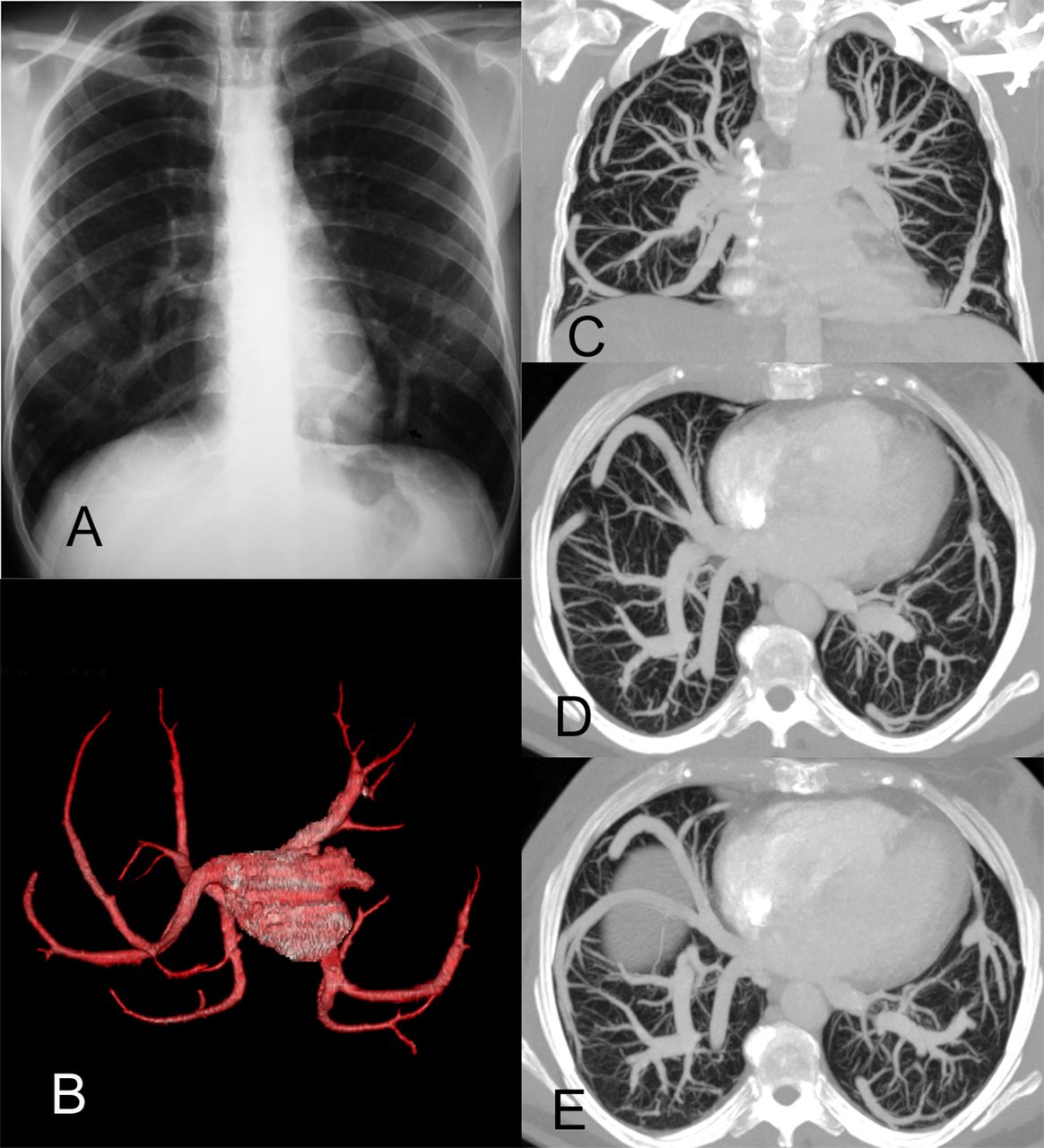
A 56-year-old woman underwent chest x-ray for evaluation of a pelvic tumour. The x-ray showed a unique pattern of bilateral, anomalous, curvilinear vessels (figure 1A). CT with volume-rendered reconstruction (figure 1B), coronal (figure 1C) and axial (figure 1D,E) maximum intensity projection (MIP) images demonstrated bilateral pulmonary veins that followed an aberrant circuitous route before draining normally into the left atrium; this pattern is referred as bilateral anomalous single pulmonary veins.
Chest x-ray showed a unique pattern of bilateral, anomalous, curvilinear vessels (A). CT with coronal volume-rendered reconstructions (B) and coronal (C) and axial (D and E) MIP CT images demonstrated bilateral pulmonary veins following an aberrant circuitous route before draining normally into the left atrium. MIP, maximum intensity projection.
Although anomalous pulmonary veins are a benign malformation with normal venous return, usually asymptomatic, this condition must be distinguished from anomalous pulmonary venous connections (sometimes associated with scimitar syndrome), which result in a left-to-right shunt.1 ,2
Footnotes
-
Contributors All authors designed the study, analysed the samples and the data, wrote the manuscript, and revised and checked it.
-
Competing interests None.
-
Patient consent Obtained.
-
Provenance and peer review Not commissioned; externally peer reviewed.