Article Text

Statistics from Altmetric.com
- CT spiral
- benign symmetrical lipomatosis
- mediastinal disease
- pleural disease
- pneumonia
- tuberculosis
- ARDS
- empyema
A 34-year-old woman presented to the chest physician with progressively increasing shortness of breath. She was non-diabetic, non-hypertensive, non-obese without any other co-morbid illnesses. There was no history of prolonged drug intake of any sort. Other associated diseases like Cushing's syndrome were excluded. A chest radiograph (postero-anterior view) revealed a veil-like opacity in the mediastinum obscuring the cardiac borders (figure 1). To further evaluate the nature of mediastinal pathology, a chest CT was done. The CT showed gross, bilaterally symmetrical masses of adipose tissue in the mediastinum causing segmental atelectasis of the lungs (figure 2). The fatty tissue extended from the superior mediastinum into the anterior mediastinum up to the diaphragm. It exhibited a range of attenuation values from −155 to −110 Hounsfield units. The diagnosis of extensive mediastinal lipomatosis was established, which was proven by a biopsy from the mass. In this condition, mature adipose tissue is deposited symmetrically in the mediastinum.1 The mediastinal lipomatosis in this case is idiopathic.2
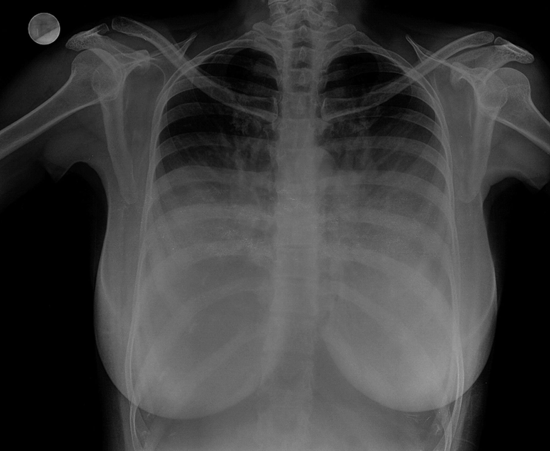
Chest radiograph (posteroanterior view) showing extensive veil-like mediastinal widening obscuring the cardiac borders.

(A, B) Axial non-contrast CT showing fatty symmetrical masses in the mediastinum.
Learning points
Gross symmetric mediastinal widening in the absence of trauma can be caused by benign entities like lipomatosis.
CT and MRI are usually sufficient to establish a diagnosis.
Footnotes
Competing interests None.
Patient consent Obtained.
Ethics approval The ethics approval was provided by institutional ethics committeee.
Provenance and peer review Not commissioned; internally peer reviewed.