Article Text

Statistics from Altmetric.com
- Bronchiectasis
- non-invasive ventilation
- sarcoidosis
- lung cancer
- respiratory infection
- tuberculosis
- COPD mechanisms
- asthma
- asthma mechanisms
- COPD exacerbations
- cough/mechanisms/pharmacology
- COPD epidemiology
- COPD pathology
- COPD pharmacology
- emphysema
- health economist
- bronchoscopy
- rare lung diseases
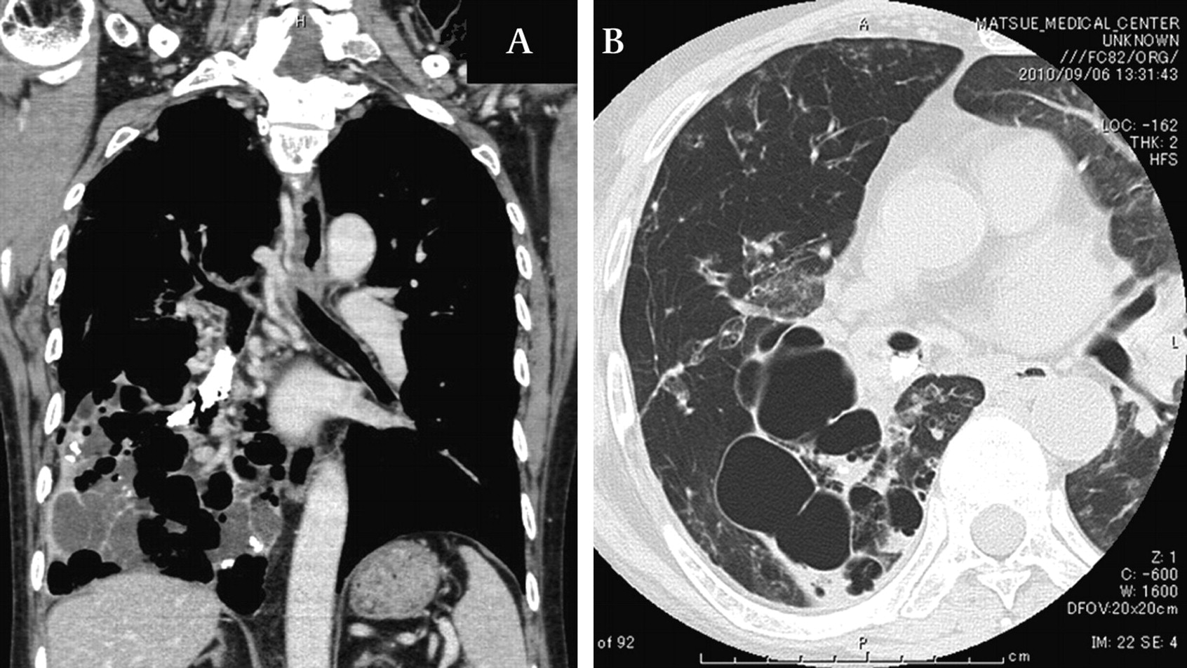
We present an unusual type of broncholithiasis complicated with cystic bronchiectasis. Chest CT scans showed cystic bronchiectasis with fluid collection in the right lower lobe and calcification was also detected in the right lower bronchus (figure 1A,B). Bronchoscopy showed a ‘coral’ broncholith arising from the right lower bronchus (figure 2A,B).

(A) Coronal section of chest CT. Calcified lesion is seen in the lower bronchus. Cystic lesion is evident in the right lower lobe. (B) Chest CT showing cystic bronchiectasis in the right lower lobe.

(A,B) Bronchoscopy showing ‘coral’ broncholith arising from the right lower bronchus.
Broncholithiasis is commonly caused by erosion and extrusion of a calcified adjacent lymph node into the bronchial lumen, a finding usually associated with tuberculosis or histoplasmosis.1 In the present case, cystic bronchiectasis might be associated with broncholith formation.
Learning points
This case is the first report of the ‘coral’ broncholith.
Cystic bronchiectasis might be associated with the broncholith formation.
Reference
Footnotes
Competing interests None.
Patient consent Obtained.
Provenance and peer review Not commissioned; externally peer reviewed.