Article Text

Statistics from Altmetric.com
S62 ABERRANT PERIPHERAL NEUTROPHIL MIGRATION IN THE HEALTHY ELDERLY AS A POTENTIAL CAUSE OF REDUCED BACTERIAL CLEARANCE
1E. Sapey, 1N. Aaronson, 1A. Ahmad, 1H. Chalal, 2R. H. Insall, 1J. M Lord, 3R. A. Stockley. 1University of Birmingham, Birmingham, UK, 2Beatson Institute, Glasgow, UK, 3University Hospital Birmingham NHS Foundation Trust, Birmingham, UK
Pneumonia is the leading infectious cause of death in the elderly, and associated with a poor response to antimicrobial therapy. Neutrophil function declines with age, with reduced phagocytosis and superoxide production. In contrast there have been conflicting data as to whether neutrophil migration is altered during ageing. Inaccurate migration of neutrophils to a site of infection may reduce pathogen clearance and cause an increase in “by-stander” tissue damage as neutrophils use proteases to aid migration through tissue. The aim of this study was to assess whether migratory dynamics of circulating neutrophils were different in the healthy aged.
Methods Migratory parameters (including chemotaxis (velocity), chemokinesis (speed), directional changes and accuracy) were measured in neutrophils isolated from 20 healthy elderly (age >65 years) and 20 young subjects (age <35 years). Neutrophils were then incubated with a CXCR2 inhibitor in order to assess its effect on migration towards the chemokine interleukin-8 (IL-8), a ligand of CXCR2. CXCR2 receptor expression and shedding were measured by immunostaining.
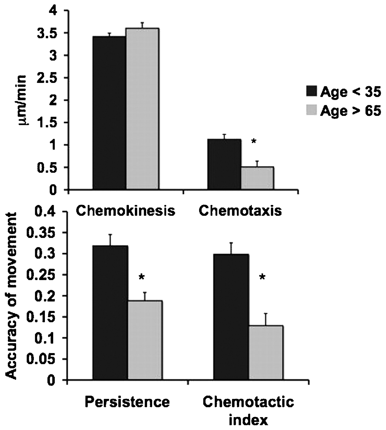
Results Neutrophils from elderly subjects migrated with the same speed (chemokinesis) but reduced velocity (chemotaxis; p<0.0001), and with reduced directional persistence and accuracy towards IL-8 (p<0.0001). Figure 1 describes differences in migration between elderly and younger subjects. Preincubating neutrophils from younger subjects with a CXCR2 antagonist reduced their velocity, persistence and accuracy to levels comparable with those from elderly subjects. There was no difference in surface expression of CXCR2; however, cells from elderly subjects shed more receptors over time (p = 0.008), suggesting increased receptor turnover with age.
Conclusions Neutrophils from elderly subjects demonstrate reduced chemotactic efficiency when migrating towards IL-8, which could reduce the efficiency of bacterial clearance during infections. The inhibition of CXCR2 signalling in younger subjects’ neutrophils produced an “old migration phenotype”, suggesting that altered chemokine receptor signalling is likely to underlie reduced chemotaxis in neutrophils with age.
The mean neutrophil migratory parameters in elderly and younger subjects (with standard error bars) during migration towards interleukin-8.
S63 THE STREPTOCOCCUS PNEUMONIAE CAPSULE PROTECTS AGAINST ALVEOLAR MACROPHAGE-MEDIATED EARLY LUNG INNATE IMMUNITY
1E. Camberlein, 1C. J. Hyams, 2J. N. Weiser, 1S. Khandavilli, 1J. M. Cohen, 1J. S. Brown. 1University College London, London, UK, 2Department of Microbiology, University of Pennsylvania, Philadelphia, USA
Introduction The polysaccharide capsule is the main Streptococcus pneumoniae virulence factor, and is the target for all existing vaccines. However, little is known about how the capsule aids bacterial immune evasion during early lung infection and why different capsular serotypes vary in their virulence. We have used mouse models to investigate the effect of the capsule on interactions with the initial phagocytic lung cell, the alveolar macrophage (AM).
Methods The effects of the capsule on the kinetics of bacterial interactions with macrophages were assessed in vitro using RAW cells and a genetically modified unencapsulated capsular serotype 4 S pneumoniae strain. Additional experiments in mice investigated capsule effects on interactions with AMs during early lung infection, using bacteria and cells recovered by bronchoalveolar lavage (BAL) for flow cytometry and microscopy assays.
Results Flow cytometry assays demonstrated that the capsule markedly inhibited both complement-dependent and complement-independent RAW cell phagocytosis of S pneumoniae. Within 15 min unencapsulated but not encapsulated bacteria were associated with RAW cells, and large differences persisted over 4 h. Similarly, within 30 min of lung infection AM association with unencapsulated (relative fluorescent intensity (RFI) 27 760±2240) was increased compared with encapsulated (RFI 7320±680) bacteria and was associated with a 2–3 log10 decrease in BAL bacterial colony-forming units (CFU) for unencapsulated bacteria by 4 h. Depletion of lung AMs using liposomal clodronate demonstrated that decreases in BAL CFU for unencapsulated bacteria were largely dependent on AMs (at 2 h, log10 CFU 5.5±0.43 in clodronate-treated mice vs 4.4±0.41 in controls), and repeated experiments using complement-deficient mice (C3−/−, C1qa−/− and Bf−/−) demonstrated that differences between unencapsulated and encapsulated bacteria were partially dependent on the classical complement pathway. Interestingly, experiments using otherwise isogenic bacteria expressing different capsular serotypes demonstrated that serotypes associated with low frequency of invasive disease were more rapidly cleared from the lungs than serotypes frequently associated with invasive disease.
Conclusion The S pneumonia capsule is vital for evasion of AM-mediated early lung immunity through inhibition of bacterial interactions with different phagocyte receptors, and differences between capsular serotypes in AM interactions may partially explain variations in virulence between S pneumoniae strains.
S64 PREVIOUS COLONISATION PROTECTS AGAINST PNEUMOCOCCAL PNEUMONIA: LEARNING FROM NATURAL IMMUNITY
1S. Khandavilli, 1E. Camberlein, 2H. Baxendale, 1J. Brown, 1J. M. Cohen. 1UCL Centre for Respiratory Research, London, UK, 2UCL Institute of Child Health, London, UK
Introduction and Objectives Existing Streptococcus pneumoniae vaccines have poor efficacy vs pneumonia in adults, and new preventative strategies are required. We have used mouse models to investigate the immunological response to S pneumoniae nasopharyngeal colonisation and its protective efficacy against subsequent pneumonia, aiming to identify natural mechanisms of protection that could be adapted to prevent S pneumoniae pneumonia in adults.
Methods Mice were colonised with wild-type (WT) or mutant D39 S pneumoniae strains, and subsequently given a lethal S pneumoniae pneumonia challenge. Disease course and immunological responses were assessed using blood, bronchoalveolar lavage (BAL) fluid and lungs recovered from infected mice for bacterial culture, microscopy, flow cytometry, ELISAs and Luminex bead assays.
Results WT S pneumoniae colonised the mouse nasopharynx for 17 days, inducing serum and BAL immunoglobulin G (IgG) against subcapsular but, surprisingly, not capsular antigens. Colonisation with the WT D39 strain protected mice against subsequent lethal pneumonia challenge (75% survival vs 25% in controls, p<0.01). Mutant strains (D39ΔD unencapsulated strain, D39Δlgt lipoprotein-deficient strain, or the structurally normal D39Δpab auxotrophic strain) were cleared more rapidly from the nasopharynx than the WT strain, induced only low levels of antibody and did not protect against subsequent pneumonia. These data suggest that the duration of colonisation and the inflammatory response is important for protective immunity to develop. During subsequent S pneumoniae pneumonia, mice previously colonised with WT bacteria had higher BAL neutrophil numbers at 4 h (2.0×10⁵/ml vs 1.1×10⁵/ml, p<0.01), and raised levels of cytokines interleukin-17 (IL-17) (78.0 pg/ml vs 17.1 pg/ml, p<0.01) and KC (68 pg/ml vs undetectable, p<0.01) compared with controls. Colonisation prevented progression from pneumonia to septicaemia (0% vs 66.7% controls, p = 0.014). Passive transfer of serum from colonised to naïve mice demonstrated that this effect was largely attributable to antibody. Additional experiments using antibody-deficient mice, CD4 cell depletion and IL-17 blockade will further characterise mechanisms of immunity to S pneumoniae pneumonia after nasopharyngeal colonisation.
Conclusions Nasopharyngeal colonisation with S pneumoniae protects against pneumonia through inducing antibodies to subcapsular antigens rather than the capsule, and possibly through a cell-mediated (Th17?) response. These data help define the parameters required for new therapeutic strategies to prevent S pneumoniae pneumonia.
S65 THE STREPTOCOCCUS PNEUMONIAE CAPSULE INHIBITS MACROPHAGE ACTIVATION THROUGH THE NF-κB AND NOT MAPK ACTIVATION PATHWAYS
C. J. Hyams, E. Camberlein, M. Noursadeghi, J. N. Weiser, J. S. Brown. UCL, London, UK
Deficiency of IRAK4- and NEMO-dependent nuclear factor-κB (NF-κB) activation results in greatly increased susceptibly to Streptococcus pneumoniae infections, demonstrating the importance of the inflammatory response for immunity to this organism. However, little is known about S pneumoniae factors that modulate the inflammatory response. The S pneumoniae polysaccharide capsule is the main virulence factor for this pathogen, but how it aids bacterial virulence is unclear. We hypothesised that the capsule could affect inflammatory responses to S pneumoniae by inhibiting macrophage–bacteria interactions. RAW 264.7 cells were used to analyse macrophage responses to a capsular serotype 4 (TIGR4) S pneumoniae strain and its unencapsulated derivative (TIGR4cps). Incubation of TIGR4cps with RAW cells caused a marked increase in the production of the inflammatory cytokine tumour necrosis factor α (TNFα) at early and late time points compared with encapsulated bacteria (at 24 h, 97.4 ng/ml SD 19.5 vs 16.9 ng/ml SD 2.6, p<0.002). Quantitative confocal immunofluorescence assays demonstrated that the capsule prevented RelA NF-κB translocation to the nucleus (analysis of variance (ANOVA), p<0.001). Capsule effects on TNFα persisted even when bacteria were not opsonised with complement. Immunoblots of IκBα degradation and phosphorylation of p38 and extracellular signal-regulated kinase (ERK) 1/2 were used to analyse the intracellular signalling pathways involved in capsule-dependent effects on the macrophage inflammatory response. The TIGR4cps strain induced greater IκBα degradation (ANOVA, p<0.001) than the encapsulated strain. However, interestingly, although both p38 and ERK1/2 were phosphorylated after incubation of RAWs with S pneumoniae, there were no significant differences in the response to unencapsulated and encapsulated bacteria for these pathways, suggesting that the capsule specifically inhibits the IκBα pathway. Overall, these data demonstrate that the capsule inhibits inflammatory responses to S pneumoniae, which may contribute to its effects on virulence. To investigate this possibility further, the inflammatory responses to otherwise isogenic TIGR4 S pneumoniae strains expressing different capsular serotypes were investigated. Incubation of RAW cells with two serotypes associated with a low frequency of invasive disease induced a larger increase in TNFα than incubation with two serotypes frequently associated with invasive disease, suggesting that differences in virulence between strains could partially depend on the ability of different capsular serotypes to mask the inflammatory response to S pneumoniae.
S66 SUBVERSION OF NEUTROPHIL APOPTOSIS BY STAPHYLOCOCCUS AUREUS: CONSEQUENCES FOR BACTERIAL PATHOGENESIS AND HOST DEFENCE
1S. Anwar, 1I. Sabroe, 2S. J. Foster, 1M. K. Whyte. 1University of Sheffield School of Medicine and Biomedical Sciences, Sheffield, UK, 2Department of Molecular Biology and Biotechnology, Sheffield, UK
Introduction Staphylococcus aureus is a major, opportunistic pathogen responsible for severe invasive infections. Since influenza virus increases the risk of staphylococcal pneumonia and S aureus is the most common cause of ventilator-associated pneumonia, it is of particular concern during the influenza pandemic. Polymorphonuclear neutrophils (PMNs) kill phagocytosed pathogens and then undergo apoptosis to limit tissue injury. In contrast, PMN death by necrosis is proinflammatory. There is increasing evidence that S aureus can cause PMN lysis and thus avoid clearance. S aureus can also indirectly influence PMNs via enterotoxin-mediated cytokine release from mononuclear cells. We aimed to characterise and quantify S aureus-mediated PMN death.
Methods S aureus SH1000 were co-cultured with ex vivo human PMNs isolated from venous blood. Apoptosis and necrosis were determined morphologically by cytospin, by flow cytometry (Annexin V/To-Pro-3) and time-lapse microscopy (Annexin V/propidium iodide). Absolute PMN counts were determined using Countbright beads.
Findings S aureus is a potent inducer of PMN necrosis which is rapid, primary and direct. Similar findings are observed with clinical methicillin-susceptible S aureus (MSSA) and hospital-acquired methicillin-resistant S aureus (HA-MRSA) strains. Necrosis occurs without evidence of prior apoptosis and is independent of caspase activation and of contaminating mononuclear cells. Both infected and non-infected PMNs are killed. Co-culture or bacterial conditioned media induce PMN necrosis, implicating a soluble bacterial virulence factor. Monocytes and macrophages are also susceptible. Bacterial replication is not limited by PMN necrosis. PMN death is attenuated by S aureus mutants of the saeR and sarA loci (global virulence regulators), but not by an agr (quorum-sensing regulator) mutant. Using a candidate approach with isogenic mutants, α-, β-, γ- and δ-haemolysins have been excluded as causative virulence factors. Current work aims to purify the virulence factor(s) using fast protein liquid chromatography (FPLC) and proteomic approaches.
Conclusions These findings demonstrate the ability of S aureus to evade PMN responses by inducing massive primary necrosis, resulting in bacterial persistence. This is not attributable to principal candidate toxins investigated thus far. Rising antibiotic resistance and virulence highlights the critical need to better understand staphylococcal innate immune evasion and enable therapeutic manipulation in future.