Article Text

Statistics from Altmetric.com
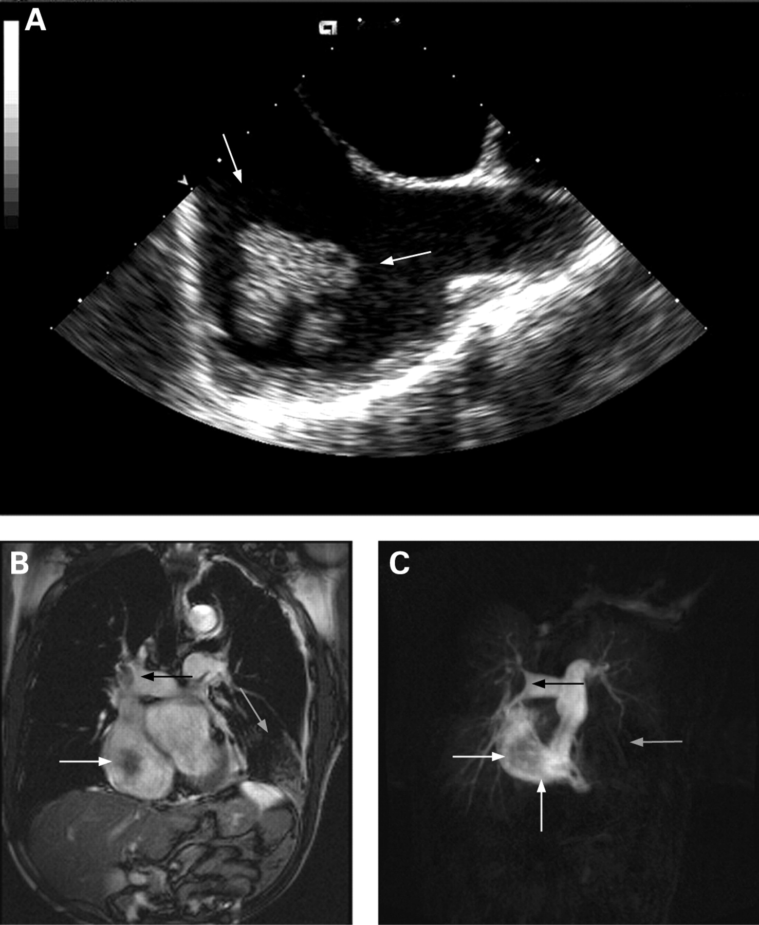
A 70-year-old man had an episode of dyspnoea followed by syncope 2 months after a stroke. Deep venous thrombosis was found in his right lower extremity by venous Doppler ultrasound. Transoesophageal echocardiography revealed a 33×38 mm highly mobile mass floating in his right atrium with no attachment to any atrium structure (fig 1A). The right heart chambers were dilated with mild tricuspid valve regurgitation and a systolic pulmonary artery pressure of 54 mm Hg. Cine magnetic resonance imaging showed a low-signal free-floating mass within the right atrium and a large thrombus in the right main pulmonary artery in the same view (fig 1B). No perfusion to the left lower lobe was observed in the perfusion view (fig 1C). Emergency thromboembolectomy was performed. The diagnosis of thrombus was confirmed by histological examination. Two years later an echocardiogram showed a normal heart structure and function.
(A) Transoesophageal echocardiogram showing a large thrombus (arrows) floating in the right atrium with no attachment to any heart chamber structure. (B) Cine magnetic resonance imaging (MRI) showing a low-signal floating mass (white arrow) in the right atrium and a large thromboembolus (black arrow) in the right main pulmonary artery. Left lower lobe consolidation can be also noted (grey arrow). (C) Perfusion view of MRI showing the floating right atrial thrombus (white arrows) and right main pulmonary thromboemboli with no perfusion to left lower lobe (grey arrow).
This case illustrates well the potential life-threatening nature of free-floating right atrial thrombus, and suggests that prompt intervention is merited whenever these lesions are detected. However, the management strategy remains controversial.1 2 Although thrombolysis is advocated first, surgical thromboembolectomy remains the classic treatment, particularly for cases in whom thrombolysis is contraindicated or if thrombolysis is ineffective. Anticoagulation with heparin is more an antithrombotic than a lytic therapy. It is inappropriate as the sole treatment of an impending pulmonary embolism.
Learning points
Nearly 100% of free-floating right heart thrombi are associated with the presence of pulmonary embolism.
Cine magnetic resonance imaging is an alternative modality for the diagnosis of right heart free-floating thrombus and pulmonary embolism.
Surgical thromboembolectomy remains the classic treatment.
Footnotes
Competing interests: None.
Patient consent: Obtained.
Linked Articles
- Airwaves