Article Text

Download PDFPDF

Is this really pneumothorax?
Free
  1. M P Singh,
  2. J Armstrong,
  3. M A Murphy,
  4. J Coucher,
  5. B Ong
  1. QE II Jubilee Hospital, Brisbane, Australia
  1. M P Singh, QE II Jubilee Hospital, Cnr Kessels and Troughton Roads, Coopers Plains, Brisbane, QLD 4108, Australia; maninder.singh{at}health.qld.gov.au

Statistics from Altmetric.com

Request Permissions

If you wish to reuse any or all of this article please use the link below which will take you to the Copyright Clearance Center’s RightsLink service. You will be able to get a quick price and instant permission to reuse the content in many different ways.

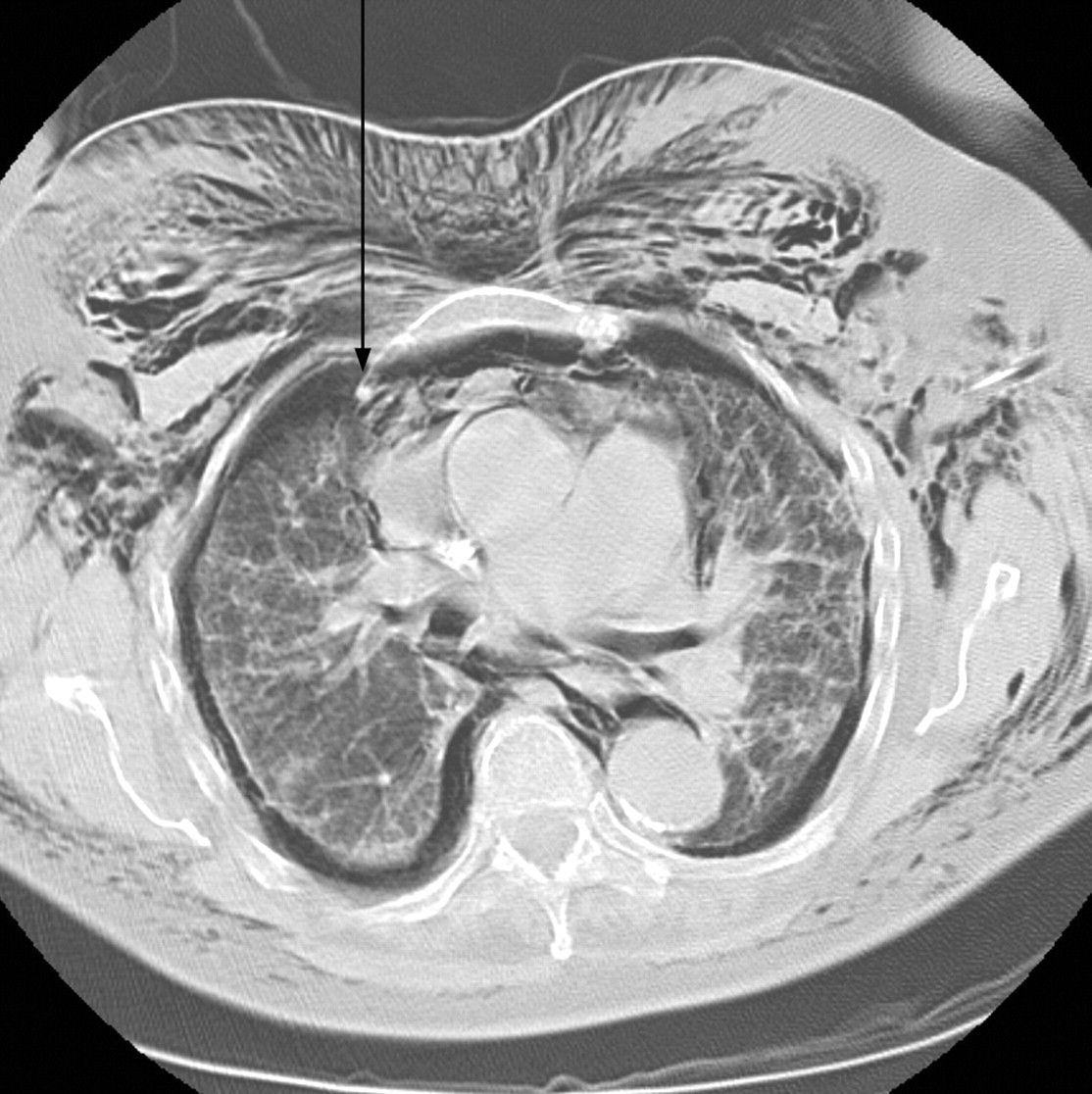
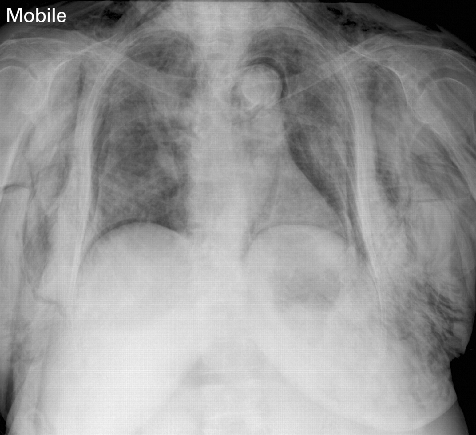
A 75-year-old female non-smoker was admitted for investigation of suspected interstitial lung disease. A left lower lobe transbronchial lung biopsy was performed, complicated by suspected bilateral pneumothoraces, pneumomediastinum and surgical emphysema (fig 1). Bilateral intercostal catheters (ICCs) were inserted. In view of the ongoing air leak and worsening surgical emphysema, a CT chest was undertaken. This was suspicious of extrapleural air leak. Following cardiothoracic consultation, video assisted thoracic surgery was performed which confirmed obliterated pleural spaces bilaterally with extrapleural air leak, as shown in fig 2. Bilateral ICCs were inserted into the air filled extrapleural space with dramatic reduction in surgical emphysema. The patient declined further invasive investigations.

Figure 1 Post bronchoscopy radiograph at 11 h demonstrates extensive subcutaneous gas. Lucency outlining the aortic knuckle and left cardiac border is consistent with pneumomediastinum. There are bilateral pleural based lucencies mimicking bilateral pneumothoraces.
Figure 2 CT chest performed 2 days post bronchoscopy shows bilateral extrapleural air collection with right internal mammary artery traversing the collection (arrow), and multiple fibrous septae within the collection, and extensive surgical emphysema. The extrapleural air collections communicate with each other anteriorly and there is no anterior junction line identifiable.

Learning points

  • Physicians caring for patients following transbronchial lung biopsy should be aware of the rare complication of bilateral extrapleural air leak mimicking pneumothoraces.1 2

  • A communication between pleural spaces may be present in patients without a history of mediastinal surgery or sternal mediastinotomy.

REFERENCES

View Abstract

Footnotes

  • Competing interests: None.

  • Patient consent: Obtained.

Linked Articles

  • Airwaves
    Wisia Wedzicha