Article Text

Statistics from Altmetric.com
From the question on page 210
This case describes an extremely rare condition of extramedullary plasmacytoma (EMP) hidden in the middle media-stinum, giving no systemic signs but causing severe central airway narrowing detectable by the pattern of the flow-volume loop. The constant expiratory and inspiratory flow limitation is consistent with severe fixed narrowing of the central airway.1 Accurate analysis of the flow-volume curve pattern at the onset of symptoms would have given rise to a suspicion of tracheal compression several months earlier.
Fixed intrathoracic airway obstruction is usually due to intramural infiltration (post-endotracheal intubation, recurrent polychondritis, primitive tracheobronchial neoplasia and amyloidosis) or to extrinsic compression (intrathoracic goitre, thymoma, lymphoma).2 Mediastinal localisation of plasma-cytoma is very uncommon, usually presenting as a large mass visible on the chest radiograph.3 In this case, EMP was hidden in the middle mediastinum and could only be detected on the chest CT scan.
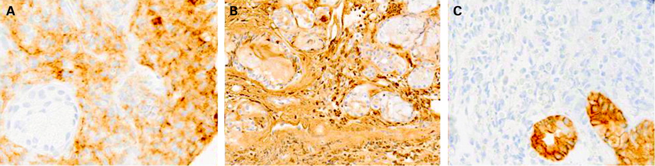
Transbronchial biopsy was consistent with IgG lambda plasmacytoma. Multiple Russell bodies on PAS staining were indicative of cytoplasmic inclusions of immunoglobulins. The results of immunohistochemical staining are shown in fig 1.
EMP is a plasma cell neoplasm of soft tissue without bone marrow involvement or other systemic characteristics of multiple myeloma, representing about 3% of all plasma cell neoplasms.4 It can be differentiated from reactive plasmacytoma and plasma cell granuloma or lymphoma (MALT, marginal and immunoblastic) by the expression of specific cell surface markers.5
This case suggests that the pattern of the flow-volume loop may give a hint of central airways narrowing caused by hidden masses not visible by traditional procedures.