Article Text

Abstract
Background: A study was undertaken to determine if quantitative CT estimates of lung parenchymal overinflation and airway dimensions in smokers with a normal forced expiratory volume in 1 s (FEV1) can predict the rapid decline in FEV1 that leads to chronic obstructive pulmonary disease (COPD).
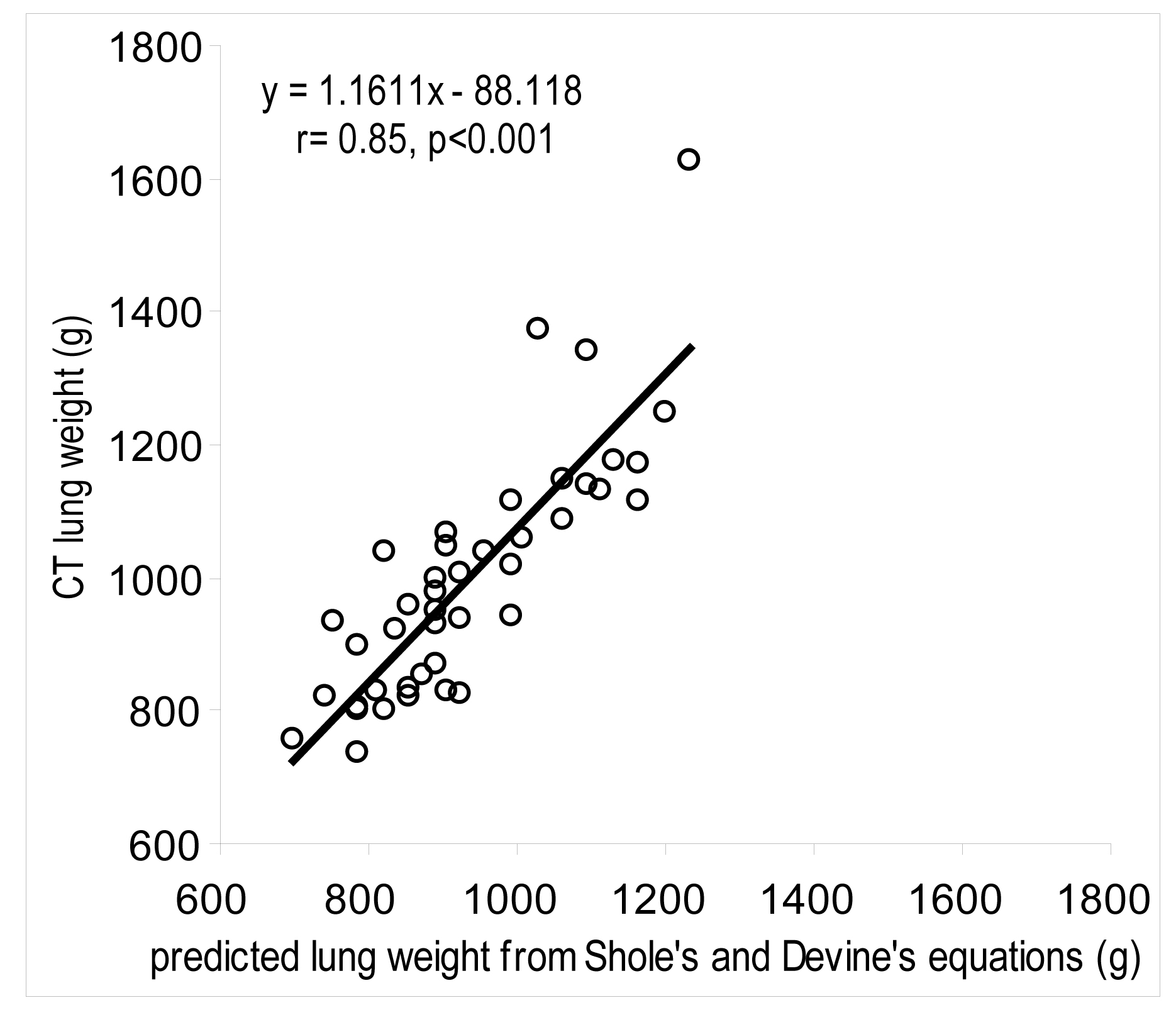
Methods: Study participants (n = 143; age 45–72 years; 54% male) were part of a lung cancer screening trial, had a smoking history of >30 pack years and a normal FEV1 and FEV1/forced vital capacity (FVC) at baseline (mean (SD) FEV1 99.4 (12.8)%, range 80.2–140.7%; mean (SD) FEV1/FVC 77.9 (4.4), range 70.0–88.0%). An inspiratory multislice CT scan was acquired for each subject at baseline. Custom software was used to measure airway lumen and wall dimensions; the percentage of the lung inflated beyond a predicted maximal lung inflation, the low attenuation lung area with an x ray attenuation lower than −950 HU and the size distribution of the overinflated lung areas and the low attenuation area were described using a cluster analysis. Multiple regression analysis was used to test the hypothesis that these CT measurements combined with other baseline characteristics might identify those who would develop an excessive annual decline in FEV1.
Results: The mean (SD) annual change in FEV1 was −2.3 (4.7)% predicted (range −23.0% to +8.3%). Multiple regression analysis revealed that the annual change in FEV1%predicted was significantly associated with baseline percentage overinflated lung area measured on quantitative CT, FEV1%predicted, FEV1/FVC and gender.
Conclusion: Quantitative CT scan evidence of overinflation of the lung predicts a rapid annual decline in FEV1 in smokers with normal FEV1.
Statistics from Altmetric.com
Supplementary materials
web only appendices
Files in this Data Supplement:
Footnotes
▸ Additional details are published online only at http://thorax.bmj.com/content/vol64/issue11
Funding HOC is a Canadian Institutes of Health Research/British Columbia Lung Association New Investigator and is also supported in part by the University of Pittsburgh COPD SCCOR NIH 1P50 HL084948 and R01 HL085096 from the National Heart, Lung and Blood Institute, National Institutes of Health, Bethesda, MD to the University of Pittsburgh. PDP is a MSFHR Distinguished scholar and the Jacob Churg Distinguished Researcher. DDS is a Canada Research Chair in COPD and a Senior Scholar with the Michael Smith Foundation for Health Research. SL is supported by NIH grant 1PO1-CA96964, U01CA96109 and NCI contract N01-CN-85188. This project was funded by a CIHR Industry partnership grant with GlaxoSmithKline.
Competing interests JCH has served as a consultant, given lectures and participated in advisory boards of several major pharmaceutical companies in the past five years. The total reimbursement for these activities is less than $20 000. PDP was the principal investigator of a Merck Frosst supported research programme to investigate gene expression in the lungs of patients who have COPD. He and collaborators have received approximately $200 000 for this project. These funds have supported the technical personnel and expendables involved in the project. He sits on an advisory board for Talecris Biotherapeutics who make anti-one antitrypsin replacement therapy. He is the principal investigator of a project funded by GlaxoSmithKline to develop CT-based algorithms to quantify emphysema and airway disease in COPD. With collaborators he has received approximately $300 000 to develop and validate these techniques. These funds have been applied solely to the research to support programmers and technicians. DDS has received research funding from GlaxoSmithKline and AstraZeneca for projects on chronic obstruction pulmonary disease. He has also received honoraria for speaking engagements for talks on COPD sponsored by these organizations. HOC received $11 000 in 2005 and $4800 in 2006 and 2007 for serving on an advisory board for GlaxoSmithKline. He is the co-investigator on two multicentre studies sponsored by GlaxoSmithKline and has received travel expenses to attend meetings related to the project. He has three contract service agreements with GlaxoSmithKline to quantify the CT scans in subjects with COPD and a service agreement with Spiration Inc to measure changes in lung volume in subjects with severe emphysema. A percentage of HOC’s salary between 2003 and 2006 (15 000 US $/year) derives from contract funds provided to a colleague PDP by GlaxoSmithKline for the development of validated methods to measure emphysema and airway disease using computed tomography. HOC is the co-investigator (with DDS) on a Canadian Institutes of Health-Industry (Wyeth) partnership grant. There is no financial relationship between any industry and the current study. RY, JCW, YN, SL and AMMcW have no competing interests in the content of this paper.
Ethics approval The University of British Columbia Clinical Ethics Review board approved the study and all subjects provided informed written consent for the use of all materials and data.
Provenance and Peer review Not commissioned; externally peer reviewed.