Article Text

Statistics from Altmetric.com
A 29-year-old woman, a known case of tuberous sclerosis, presented to our hospital with a 1-day history of multiple episodes of convulsions. This was associated with vague chest and abdominal pain. CT scans of the brain showed multiple calcific foci in the gray-white matter junctions and in the periventricular region. CT of the abdomen revealed liver hamartomas and renal angiomyolipoma.
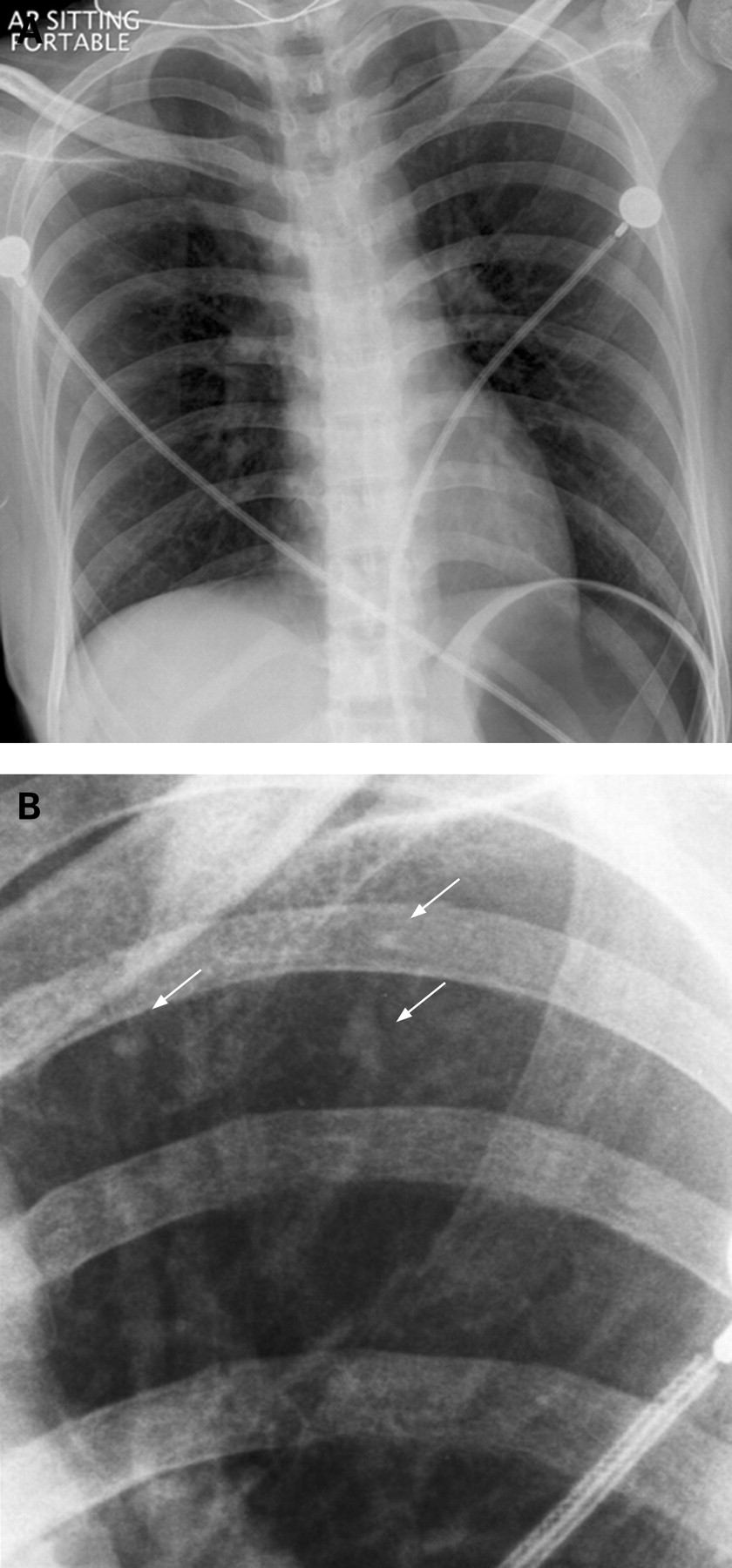
CT scans of the thorax showed multiple, tiny, randomly distributed, nodular densities of 3–10 mm in size and numerous miliary nodules 1–3 mm in both lung fields compatible with multifocal micronodular pneumocyte hyperplasia. They were more predominant in the lung periphery and the upper lobes. Few tiny simple cysts were also noted. Plain chest radiographs revealed bilateral diffuse fine nodular opacities (figs 1 and 2).


Multifocal micronodular pneumocyte hyperplasia is a recently described pulmonary manifestation of tuberous sclerosis. First described by Popper et al in 1991, there are only slightly more than 30 cases reported in the literature since. These lesions are benign hamartomatous proliferations of type II pneumocytes along alveolar septa that exhibit fibrous thickening, increased elastic fibres, and aggregated alveolar macrophages.1 2 It is usually with limited clinical significance and no known malignant potential.2
Learning points
-
Multifocal pneumocyte hyperplasia is a pulmonary manifestation of tuberous sclerosis complex that may co-exist with other lung lesions like lymphangiomatosis.
-
Multifocal pneumocyte hyperplasia should be considered, when CT revealed multiple randomly distributed pulmonary micronodules, with lung periphery and upper lobes predominance, in patients with known tuberous sclerosis complex.