Article Text

Statistics from Altmetric.com
Pleural endometriosis typically presents in young women within 72 h of onset of menstruation as cata-menial pneumothorax, haemothorax or both. A 43-year-old multiparous woman with a history of pelvic endometriosis and myocardial infarction presented with acute onset of right hemithorax pain and dyspnoea coinciding with dysmenorrhoeic menstruation. A non-contrast enhanced chest CT scan (fig 1) showed a right haemopneumothorax with floating clots within the fluid, which was drained. A video-assisted thoracoscopy (VATS) guided random pleural biopsy sample was negative. Magnetic resonance imaging (MRI) of the chest coinciding with menses 4 months later showed an oval-shaped lesion with homogeneous high signal intensity in T1- and T2-weighted images in the right posterior costodiaphragmatic recess (fig 2A, B). Owing to the relative contraindication for hormone therapy related to the coronary artery disease, the patient underwent a hysterectomy and oophorectomy for treatment of endometriosis. Six months later she was free of thoracic symptoms and an MRI scan showed the disappearance of the right pleural abnormality.

Non-contrast enhanced chest CT scan at presentation showing (A) right haemopneumothorax in the lung parenchyma and (B) evidence of slightly hyperdense floating clots (arrows) within the pleural fluid.

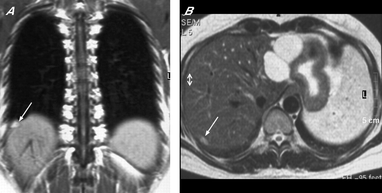
Non-contrast enhanced MRI scan of the chest and upper abdomen showing an oval-shaped well-defined lesion in the right posterior costodiaphragmatic recess abutting the pleura. The lesion (arrow) exhibits homogeneous high signal intensity in (A) the coronal T1-weighted spin echo image and (B) the axial T2-weighted fast spin echo images without fat suppression.
The diagnosis of thoracic endometriosis is usually based on clinical grounds but is often delayed. Cytological examination of the pleural fluid is usually not helpful and VATS is often negative.1,2 Although no pathological proof was available, the diagnosis of pleural endometriosis in our case was supported by the typical clinical presentation, the history of pelvic endometriosis, the MRI features distinctive of pleural endometriosis3 and the disappearance of the pleural MRI finding after surgical treatment of endometriosis.
Learning points
-
VATS is often non-diagnostic in patients with catamenial haemo-thorax or haemopneumothorax.
-
The MRI appearance of a pleural-based lesion exhibiting homo-geneous high signal in T1- and T2-weighted images is distinctive of pleural endometriosis.
-
MRI after drainage of the pleural effusion and coinciding with menses could support the diagnosis of pleural endometriosis in women with catamenial haemothorax or haemopneumothorax.