Article Text

Statistics from Altmetric.com
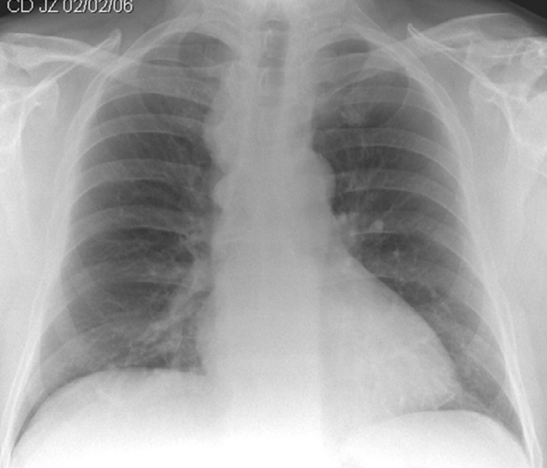
A 59-year-old builder presented to the accident and emergency department with a 4-week history of intermittent central chest pain. He was a lifelong non-smoker, had no asbestos or tuberculosis exposures, and had no cardiac risk factors. The patient’s pain settled with simple analgesia. Chest x ray showed a widening of the right paratracheal stripe consistent with right paratracheal lymphadenopathy (fig 1). Reassuringly, the computed tomography scan revealed prominent thoracic vertebral osteophytes (figs 2 and 3).

Chest x ray showing widening of the right paratracheal stripe consistent with right paratracheal lymphadenopathy.

Computed tomography scan showing prominent thoracic vertebral osteophytes.

A case of osteophytes resembling paratracheal lymphadenopathy.
Cervical osteophytes have been reported to be associated with respiratory compromise, airway obstruction and dysphagia. Extra spinal manifestations of thoracic osteophytes have been reported infrequently, but single large osteophytes have been associated with bronchial obstruction and recurrent infection.
This case demonstrates that multiple thoracic osteophytes can resemble an anterior mediastinal mass on chest x ray and should be part of the differential diagnosis. To the authors’ knowledge, this is the first reported case of osteophytes resembling paratracheal lymphadenopathy.