Article Text

Statistics from Altmetric.com
A chest radiograph performed on an asymptomatic 47 year old man under investigation for hypertension was suspicious of a retrocardiac mass. Contrast enhanced computed tomographic (CT) scanning demonstrated a large low attenuation mass encasing the oesophagus. It extended from the carina to the diaphragm and measured 15 × 7 cm in diameter (fig 1). The air space consolidation and small pleural effusion at the base of the left lung were judged incidental findings as they had completely resolved 1 week later when a CT guided biopsy was attempted. A barium swallow showed that the oesophagus was of abnormal calibre but emptied promptly. Video assisted thoracoscopic biopsy was undertaken.

CT scan showing a large retrocardiac mass encasing the oesophagus.
Learning points
-
The symptoms caused by a mass are not always correlated with its location and size.
-
The posterior mediastinum is a potential source of pathology for the respiratory physician.
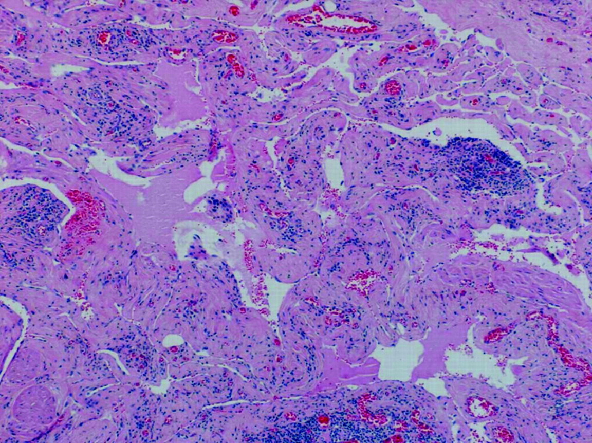
Histological examination (fig 2) showed a part cystic and part solid mass with elements of oesophageal smooth muscle and serosal surface. Ectatic vascular spaces with flat inconspicuous endothelial linings gave it a “sponge-like” appearance. Some of the spaces contained lymph and others blood. It was decided that these malformations were benign and best classified as a lymphangioma.

Thoracoscopic biopsy specimen showing ectatic vascular spaces, some containing lymph and others blood.
Oesophageal lymphangioma is an exceptionally rare cause of a mediastinal mass in adults. It is a benign proliferation of lymphatic vessels and sacs that can grow in an infiltrative fashion.1 This is the largest described in the literature.2 Complications are usually due to mass effect. Treatment is either surgical resection or conservative. Given his lack of symptoms, this patient elected for the latter.