Article Text

Abstract
Background: Mesothelioma is the most commonly occurring primary pleural neoplasm. Several studies have documented an increase in the incidence of this malignancy during the last decades. Although the association between asbestos exposure and development of mesothelioma is generally accepted, the exact mechanism of carcinogenesis is unknown. Recently, Simian virus 40 large T antigen (SV40 Tag) expression has been detected in pleural mesothelioma. The ability of SV40 oncoproteins to inactivate p53 and retinoblastoma tumour suppressor proteins has been proposed as an important step in the pathogenesis of human mesothelioma.
Methods: To obtain a better understanding of the molecular mechanisms of the pathogenesis of mesothelioma, the expression of the cell cycle inhibitor p21WAF1/CIP1 (p21), a downstream target of p53, was evaluated immunohistochemically in a group of 29 mesothelioma specimens already characterised for the presence of SV40 Tag sequences.
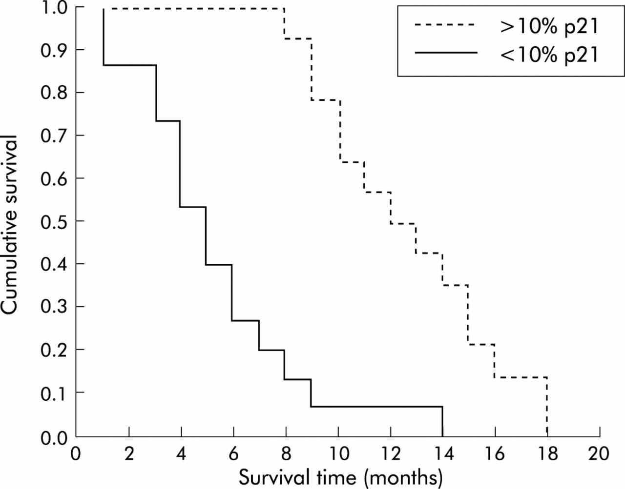
Results: Statistical analysis did not reveal any correlation between p21 expression and histopathological type of mesothelioma using the κ2 test (p=0.577). A significant positive relationship was found between p21 expression level and the patients' overall survival according to the Kaplan-Meier survival curves and using a log rank test (median difference in survival 7 months, 95% CI 4.8 to 9.9; p<0.001).
Conclusions: Determination of p21 expression bears a prognostic significance in patients affected with mesothelioma, further underlining the role of SV40 in the pathogenesis of malignant pleural mesothelioma.
- mesothelioma
- p21WAF1
- CIP1
- immunohistochemistry
- SV40 large T antigen
Statistics from Altmetric.com
Although mesothelioma is the most important primary pleural neoplasm, it accounts for less than 1% of all cancer deaths in the world.1 However, several studies have documented an apparent increase in the incidence of malignant mesothelioma during the last decades. The annual incidence of this neoplasm in the United States is estimated to be approximately 2000–3000 new cases per year.1 Even though the association of exposure to asbestos and the development of mesothelioma is generally accepted,2,3 the exact mechanism whereby asbestos induces the mesothelioma is unknown. Moreover, in the United States about 20% of these tumours are not related to asbestos exposure and less than 10% of those with high exposure to asbestos develop mesothelioma.3,4
Recent studies have proposed a role for Simian virus 40 large T antigen (SV40-Tag) in the pathogenesis of human mesothelioma,5 and multi-institutional analysis confirmed the presence of SV40-Tag sequences in a significant percentage of cases.6–9 Consistent with the possible role of SV40 oncoproteins in the pathogenesis of mesothelioma, there are observations that mutations involving p53 and the retinoblastoma (RB) gene family are extremely rare in these neoplasms,10,11 and that SV40-Tag isolated from mesotheliomas is able to bind to p53 and to the RB family proteins.12,13 Most mesotheliomas harbour CDKN2 (p16) and ARF mutations.14,15 These mutations are able to abrogate completely RB mediated growth constrains16 but not to abrogate p53 function.17 Hence, it has been proposed that p53 is the most important target of SV40 oncoproteins in mesothelioma.15
It has recently been shown that the promoter of the Cdk inhibitor p21WAF1/CIP1 (p21) contains two elements recognisable by p53 and that binding of wild type p53 to these regions upregulates p21 transcription.18 p21 is therefore considered a downstream target of p53. We have investigated the expression of p21 in a group of mesothelioma specimens already characterised for the expression of p53 and for the presence of SV40 sequences.12 We developed an immunohistochemical assay which enables us to estimate p21 expression in formalin fixed and paraffin embedded mesothelioma specimens. The aim of the study was to establish the frequency and level of p21 expression in human mesotheliomas and to correlate the results with patient survival.
METHODS
Patients
Tissues from 29 malignant mesothelioma specimens (16 epithelial, six sarcomatoid, and seven mixed mesotheliomas) obtained from open biopsy specimens or pleurectomies were evaluated. All the patients were diagnosed and treated at the Second University of Naples from 1980 to 1993. Survival data were collected from hospital charts and from periodic interviews with patients and their relatives. Two subjects who died of causes other than mesothelioma during the follow up period were excluded from the study. Only in three cases did the clinical history of the patients demonstrate a clear exposure to asbestos. The clinical and histopathological data of the patients are listed in table 1.
Correlation between p21 and histology with survival in patients with mesothelioma
All the specimens were already characterised for the presence of SV40 sequences.12
Histological examination
The formalin fixed, paraffin embedded samples were cut into sections of 5 μm thickness and stained with haematoxylin and eosin. The histological diagnosis was re-examined. In addition, the most representative blocks were selected to be cut into new 5 μm thick sections for immunohistochemical studies.
Immunohistochemistry
Sections 5 μm thick were cut from each specimen, mounted on glass, and dried overnight at 37°C. All sections were then deparaffinised in xylene, rehydrated through a graded alcohol series, and washed in phosphate buffered saline (PBS). PBS was used for all subsequent washes and for antibody dilution. Endogenous peroxidase activity was blocked by 5% hydrogen peroxide. The primary monoclonal antibody for p21 (sc-6246, Santa Cruz Biotechnology, CA, USA) was applied at room temperature for 1 hour at a dilution of 1:100. The optimal working dilution was defined on the basis of a titration experiment. The sections were then immunostained with streptavidin-biotin (Dako, Carpintera, CA, USA) using 3-amino-9-ethylcarbazide (AEC) as the final chromogen and haematoxylin as the nuclear counterstain. Negative controls for each tissue section were prepared by leaving out the primary antibody. A positive control was run with each set of slides. All samples were processed under the same conditions.
Scoring and quantification of immunoreactivity
Three observers (AB, RR, and FB) estimated the staining pattern of p21 separately and scored each specimen for the percentage of positive nuclei (≤10% and >10% of cells expressing p21). The level of concordance, expressed as the percentage of agreement between the observers, was 90% (27 of 30 specimens). In the remaining specimens the score was obtained after collegial revision and agreement.
Statistical analyses
The cumulative probability of death was calculated for each group according to the Kaplan-Meier method and compared using log rank test. To evaluate the influence on survival of p21 expression the population was divided into two groups: ≤10% and >10% of cells expressing p21. The influence of histological type on survival was evaluated by dividing the specimens into epithelial, sarcomatoid, and mixed types. Correlation between the rate of expression of p21 and histological type was performed using a κ2 test, grouping mesotheliomas as epithelial or non-epithelial. There was no need to perform multivariate analysis to estimate the influence of predictive factors. A p value of ≤0.05 in a two tailed test was considered significant. SPSS software Version 9.00 (SPSS, Chicago, IL, USA) was used for statistical analysis.
RESULTS
Twenty nine specimens (16 epithelial, six sarcomatoid, and seven mixed mesotheliomas) were evaluated. All were positive for the presence of SV40 sequences.12 Immunoreactivity for p21 was found in both normal and neoplastic tissues. p21 immunostaining was always nuclear, with a low to absent background (fig 1). The number of positive cells varied in different specimens (table 1). Fifteen of the 29 specimens had ≤10% positivity for p21 expression; the remaining 14 specimens had a higher percentage of positive cells. No significant correlation was found between p21 expression and histological type using the κ2 test (p=0.577). The median survival time was 5 months for the group with ≤10% positive cells and 12 months for those with >10% positive cells (median difference 7.0 months, 95% CI 4.8 to 9.9).
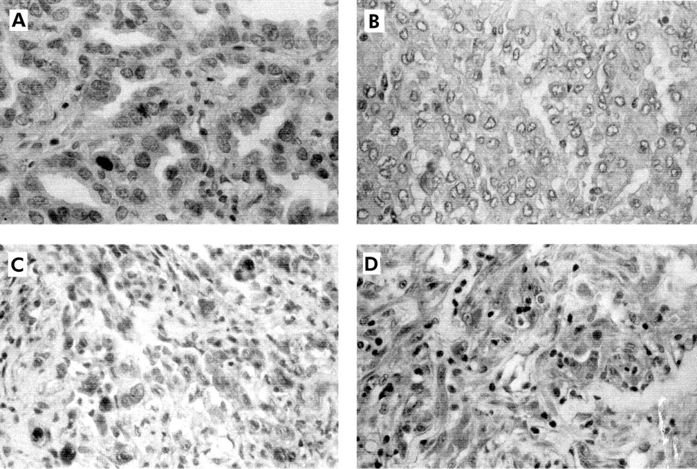
(A) Epithelial mesothelioma with ≤10% of cells positive for p21. (B) Epithelial mesothelioma negative for p21 staining. (C) Mixed mesothelioma with ≤10% of cells positive for p21. (D) Sarcomatoid mesothelioma with >10% of cells positive for p21 staining. All sections were stained as described in the text. Original magnification ×400 for all sections.
A positive relationship was found between the level of p21 expression and overall survival. To support the validity of these data, survival curves were constructed using Kaplan-Meier survival analysis (fig 2). The cumulative probability of death was calculated for each group according to the Kaplan-Meier method and compared using the log rank test (p<0.001).

Cumulative probability of death in the two mesothelioma groups (≤10% and >10% of p21 positive cells) according to the Kaplan-Meier method.
DISCUSSION
In this study we found p21 protein expressed both in normal and in neoplastic tissue. Immunohistochemical analysis was chosen for this investigation because it has the advantage of allowing visualisation of the staining pattern of each section, enabling non-neoplastic elements such as stromal, endothelial, and inflammatory cells (which always affect nucleic acid based approaches) to be excluded. Moreover, recent studies have shown that p21 protein and RNA expression are strictly correlated.19
p21 is generally considered a downstream target of p53.18 Nevertheless, since a basal level of p21 can be found in p53–/– cells, it appears that p21 can also be induced by pathways independent of p53.20–22 The p21 pathway inhibits DNA replication by interacting with PCNA, but it has also been shown that this same inhibitory interaction with p21 does not affect PCNA dependent DNA repair.23 It has recently been proposed that the SV40-Tag detected in mesothelioma specimens could play a part in the pathogenesis of this malignancy by impairing the function of several tumour suppressor and growth suppressor proteins such as p53 and the RB gene family.12,13 However, the relevance of SV40 to the pathogenesis of mesothelioma is not universally accepted, and there is epidemiological evidence that does not support a link between SV40 infection and mesothelioma.24 Moreover, most mesotheliomas retain mutations involving the CDKN2/ARF locus,14,15 impairing the functional unit composed by D-type cyclins, Cdk4/6 kinases, p16 and pRb proteins which govern the G1 phase of the cell cycle.25 It is possible that the most important target of SV40 in mesothelioma tumorigenesis is the p53 pathway. Indeed, it has been shown that abrogation of SV40-Tag expression in mesothelioma cells coincides with enhanced expression of p21.15 It has also been shown that the ability of p14-ARF to reduce the growth rate of mesothelioma cells and to induce apoptosis depends on the overexpression of p21.26
These observations clearly show that p21 acts as a key element in the control of cell growth and tumorigenesis in mesothelioma cells. The importance of p21 in the acquisition of the malignant phenotype has recently been confirmed in double knock out studies showing that an increase of CDK activity in the G1 phase of the cell cycle is a critical step in tumour growth.27
We did not find any relationship between p21 expression and histological type in the mesothelioma specimens, but there was a positive relationship between the level of p21 expression and the overall survival of the patients which suggests that the p21 pathway is involved in the pathogenesis of mesothelioma. This could occur by silencing p21 through p53 inactivation by SV40 proteins and/or the weaker activation of p21 by alternative pathways. A greater reduction in p21 expression will cause greater aggression and result in a poorer prognosis.
This is the first study of which we are aware of the relationship between p21 expression and survival of patients with mesothelioma. The immunohistochemical expression of p21 could be useful as a prognostic marker for human mesothelioma and may represent a new target for a molecular approach to treatment. Further studies with a larger number of patients are needed to confirm these observations, but the results of this study offer a background to understanding the role of SV40-Tag in the pathogenesis of malignant pleural mesothelioma.
Acknowledgments
This work was supported in part by AIRC and Ministero della Sanità grants to MGP and by Second University of Naples grants to FB. AB is recipient of an FIRC fellowship. The authors thank Dr Pier Giorgio Natali (Regina Elena Institute, Rome, Italy) for his support and critical review of the manuscript.
REFERENCES
Footnotes
-
AB and AMG contributed equally to this work.