Article Text

Abstract
Background: It has been shown that treatment with a long acting β2 agonist in addition to a glucocorticoid is beneficial in the treatment of asthma. In asthma inflammatory cells, particularly eosinophils, migrate into the pulmonary tissue and airway lumen by means of adhesion molecules expressed on resident tissue cells—that is, fibroblasts—and become activated by cytokines and adhesive interactions. A study was undertaken to determine whether an interaction exists between the long acting β2 agonist formoterol and the glucocorticoid budesonide on inhibition of adhesion molecule expression, as well as chemo/cytokine production by human lung fibroblasts.
Methods: Lung fibroblasts were preincubated with therapeutically relevant drug concentrations of 10−8 M to 10−10 M. Cells were stimulated with interleukin (IL)-1β (1 or 10 U/ml) for 8 hours and supernatants were collected for measurement of GM-CSF and IL-8 concentrations. The cells were fixed and subjected to a cell surface ELISA technique to measure the expression of ICAM-1 and VCAM-1.
Results: Formoterol exerted an additive effect on the inhibition of IL-1β stimulated ICAM-1 and VCAM-1 upregulation and GM-CSF production by budesonide in concentrations of 10−9 M and above (p<0.05). IL-8 production was not influenced by formoterol.
Conclusion: Formoterol exerts an additive effect on the anti-inflammatory properties of budesonide. In vitro data support the finding that the combination of budesonide and formoterol in asthma treatment strengthens the beneficial effect of either drug alone.
- human lung fibroblasts
- adhesion molecules
- cytokines
- budesonide
- formoterol
- VCAM-1, vascular cell adhesion molecule
- ICAM, intercellular adhesion molecule
- GM-CSF, granulocyte-macrophage colony stimulating factor
- IL, interleukin
- AP-1, activator protein-1
- NfκB, nuclear factor κB
- PKA, protein kinase A
Statistics from Altmetric.com
- VCAM-1, vascular cell adhesion molecule
- ICAM, intercellular adhesion molecule
- GM-CSF, granulocyte-macrophage colony stimulating factor
- IL, interleukin
- AP-1, activator protein-1
- NfκB, nuclear factor κB
- PKA, protein kinase A
The treatment of asthma generally consists of inhaled glucocorticoids supplemented with inhaled β2 adrenoceptor agonists. Clinical studies show that the addition of the long acting β2 agonist formoterol to the glucocorticoid budesonide,1 as well as the combination of salmeterol and beclomethasone dipropionate or fluticasone propionate,2, 3 result in an improvement in asthma symptoms and lung function and a reduction in the number of exacerbations. There are reports of interactions between β2 agonists and glucocorticoids on the mechanistic level4 and in vitro studies have shown additive and synergistic effects on the inflammatory properties of resident pulmonary cells.5, 6
Glucocorticoids inhibit virtually all steps in the inflammatory response including cytokine production and adhesion molecule upregulation on a variety of cell types. For instance, budesonide and dexamethasone have been shown to inhibit ICAM-1 and VCAM-1 expression on activated epithelial and endothelial cells7, 8 and on fibroblasts.9 Glucocorticoids also inhibit GM-CSF, IL-8 and IL-6 production by epithelial cells10 and fibroblasts.11
Long acting β2 agonists are generally considered to be smooth muscle relaxants, while their anti-inflammatory properties are still a matter of debate. Their anti-inflammatory effects are indicated by an inhibitory effect on granulocyte adhesion to epithelium12 and on infiltration of inflammatory cells in the skin and lung of guinea pigs.13 Wallin and colleagues also recently reported the inhibition by formoterol of eosinophil infiltration in asthma.14 Formoterol inhibits ICAM-1 and VCAM-1 upregulation on human lung fibroblasts as induced by different cytokines.9 Salmeterol has been shown to inhibit GM-CSF production in blood mononuclear cells after Der p 1 stimulation.15
Human lung fibroblasts may be involved in the inflammatory process of asthma.16 In vitro they express adhesion molecules such as ICAM-1 and VCAM-1 and increase their expression after specific cytokine stimuli,17 thus possibly facilitating adhesion and transmigration of inflammatory cells in the lungs.18 Furthermore, lung fibroblasts produce large amounts of GM-CSF and IL-8 after stimulation by proinflammatory cytokines. These products are able to activate and/or attract eosinophils and neutrophils. The effects of the combination of inhaled budesonide and formoterol in vivo may therefore be partly achieved through modulation of lung fibroblast activation.
We have investigated the effect of formoterol on the anti-inflammatory action of budesonide reflected by inhibition of adhesion molecule (ICAM-1, VCAM-1) upregulation and cyto/chemokine (GM-CSF, IL-8) production by human lung fibroblasts.
METHODS
Fibroblast culture
Pulmonary parenchyma was obtained from bilobal lung resection material (of healthy lobe) after oncological surgery from a non-asthmatic individual. Fibroblasts were obtained using the explant technique in which fibroblasts grow from small pieces of tissue in a six-well plate in Ham's F12 medium (BioWhittaker, Verviers, Belgium) supplemented with 10% fetal calf serum (FCS) (Bodinko BV, Alkmaar, The Netherlands), 125 U/ml Na-penicillin G (Yamanouchi Pharma, Leiderdorp, The Netherlands), and 125 μg/ml streptomycin sulphate (Radiumfarma-Fisiopharma, Milano, Italy), hereafter referred to as Ham's complete medium. Fibroblasts were cultured in Ham's complete medium and passaged by trypsinisation with trypsin-EDTA (BioWhittaker) in a 1:4 ratio, grown to confluence (passage 5) in 7 days (microscopical examination) in 96-well or 24-well culture plates (Costar Europe Ltd, Badhoevedorp, The Netherlands) and used for experiments. Fibroblast characterisation was performed with antibodies against vimentin, cytokeratin, desmin, smooth muscle actin, and fibronectin using fluorescence microscopy. Fibroblast purity was more than 98%, the only contaminating cells being smooth muscle cells. Two other lung fibroblast strains derived from tissue of two different non-asthmatic individuals were also used and comparable results were achieved.
Drugs
Budesonide was obtained from a Pulmicort Turbuhaler (Astra Pharmaceutica BV, Zoetermeer, The Netherlands) and dissolved in 96% ethanol in a concentration of 10−2 M. Subsequently, solutions of 10−10 to 10−8 M budesonide (considered therapeutically relevant concentrations19) were prepared in Ham's complete medium. Formoterol fumarate dihydrate (Astra Draco AB, Lund, Sweden) was dissolved in DMSO in a concentration of 10−2 M. Working solutions (10−10 to 10−8 M) considered to be therapeutically relevant20 were prepared in Ham's complete medium. Viability of confluent fibroblasts after incubation with different concentrations of budesonide, formoterol, or the combination was assessed using trypan blue exclusion and was always more than 95%.
Inhibition of ICAM-1 and VCAM-1 expression
Confluent fibroblast layers in 96-well plates were preincubated for 45 minutes with different concentrations of budesonide and/or formoterol (10−10–10−8 M) followed by stimulation for 8 hours (providing optimal expression17) with 1 U/ml IL-1β (to mimic a chronic inflammatory environment) (Boehringer Mannheim, Mannheim, Germany) in the presence of budesonide alone, formoterol alone, or a combination of the two drugs (total volume 100 μl). Ethanol and DMSO were used as vehicle controls for budesonide and formoterol 10−8 M, respectively. Ham's complete medium was used as baseline control. In additional experiments, 1 hour before incubation of the combined drugs the cells were preincubated either with budesonide alone or formoterol alone in order to test possible divergent effects resulting from a difference in the sequence of addition. Subsequently, fibroblasts were washed twice with 200 μl cold PBS supplemented with 0.01% CaCl2 and fixed for 10 minutes in 96% ethanol at 4°C. Fibroblasts were dried on air for 30 minutes and stored at 4°C for maximally 14 days until determination of adhesion molecule expression.
Determination of ICAM-1 and VCAM-1 expression was performed using a modified CSE assay according to Piela and coworkers21 as described recently.17 Briefly, non-specific binding sites on fibroblast layers were blocked with 1% bovine serum albumin (BSA) and subsequently incubated with anti-ICAM-1, anti-VCAM-1 (R&D Systems, Abingdon, UK) or IgG1 isotype control (CLB, Amsterdam, The Netherlands) for 120 minutes at room temperature. Fibroblast layers were washed and incubated with HRP conjugated rabbit anti-mouse antibody (DAKO A/S, Glostrup, Denmark) for 30 minutes. After the second washing step, substrate solution containing o-phenylene diamine dihydrochloride (Sigma Chemical Co, St Louis, MO, USA) was added and colour development was stopped after 30 minutes by 3 M H2SO4. Absorbance at 490 nm was measured using a microplate reader.
Cytokine production
Confluent fibroblast layers in 24-well plates were preincubated for 45 minutes with different concentrations of budesonide and/or formoterol, followed by stimulation for 8 hours with 10 U/ml IL-1β (Boehringer Mannheim, Mannheim, Germany) in the presence of budesonide, formoterol, or a combination of the drugs (total volume 1 ml). Ham's complete medium was used as a control. Cell-free media were stored at –80°C for later measurement of cytokine concentrations using GM-CSF (R&D Systems) and IL-8 (CLB, Amsterdam, The Netherlands) enzyme linked immunosorbent assay (ELISA) kit, according to the manufacturer's instructions.
Data analysis
ICAM-1 and VCAM-1 expression was calculated the as mean values of optical density (OD, 490 nm) from quadruplicate determinations within one experiment after subtracting OD values of IgG1 isotype control. Coefficients of variation did not exceed 10%; outlying OD values within quadruplicates were only omitted when they exceeded 2SD values of the mean. To evaluate statistical differences the mean OD of the quadruplicate determinations representing ICAM-1 and VCAM-1 expression and GM-CSF and IL-8 concentrations in pg/ml were tested using the non-parametric Wilcoxon signed rank test for related samples and Friedman's test. Six separate experiments were performed. Differences were considered significant at p<0.05. Percentages of inhibition are presented as median values with interquartile ranges. IC50 values were calculated from the individual concentration response curves by linear regression and are presented as median values with interquartile ranges.
RESULTS
Inhibition of ICAM-1 and VCAM-1 upregulation on lung fibroblasts
IL-1β upregulated ICAM-1 and VCAM-1 expression significantly on human lung fibroblasts (table 1). Budesonide and formoterol both inhibited IL-1β induced ICAM-1 (fig 1A) and VCAM-1 (fig 1B) upregulation in a dose dependent manner, being significant at concentrations of 10−9 and 10−8 M. Inhibition by the combination of budesonide and formoterol was significantly larger than with either of them used alone in all assessed concentrations. Furthermore, inhibition of ICAM-1 and VCAM-1 upregulation occurred at a concentration of 10−10 M (16 (9–36)% and 47 (8–56)%, respectively). Formoterol had mainly an additive effect on the inhibition of IL-1β induced ICAM-1 and VCAM-1 upregulation by budesonide on lung fibroblasts. Vehicle controls for 10−8 M did not significantly influence ICAM-1 and VCAM-1 upregulation, except for significantly enhanced VCAM-1 upregulation by ethanol (budesonide vehicle control, 124 (95–176)%, fig 1B). IC50 values for budesonide, formoterol, and the combination of the two drugs are presented in table 2.
Effect of IL-1β stimulation on adhesion molecule expression and cytokine production of human lung fibroblasts
Median (interquartile range) IC50 values of budesonide, formoterol, and the combination of the two drugs on inhibition of GM-CSF production and ICAM-1 and VCAM-1 upregulation by human lung fibroblasts after IL-1β stimulation
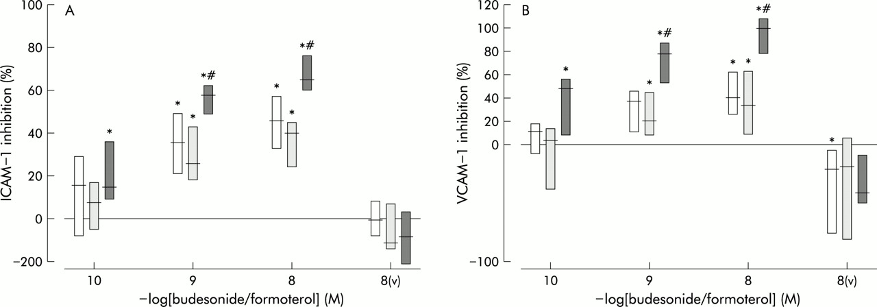
Effects of budesonide (open bars), formoterol (dark shaded bars) or both (light shaded bars) in concentrations of 10−10–10−8 M on (A) ICAM-1 and (B) VCAM-1 upregulation of human lung fibroblasts induced by IL-1β (1 U/ml). Results are presented as median percentages of inhibition with interquartile range (n=6). v=vehicle control of 10−8 M budesonide, formoterol, or both. *p<0.05 compared with expression after IL-1β stimulation; #p<0.05 compared with inhibition of budesonide and formoterol alone (additive effect).
We also assessed the effect of the combination of budesonide and formoterol when one of the two was preincubated for 1 hour before the combined drugs to determine any possible divergent effects when the sequence of addition of formoterol and budesonide was changed. No significant differences were found between additional preincubation of the separate drugs and the simultaneous incubation of budesonide and formoterol (data not shown).
Inhibition of GM-CSF and IL-8 production by lung fibroblasts
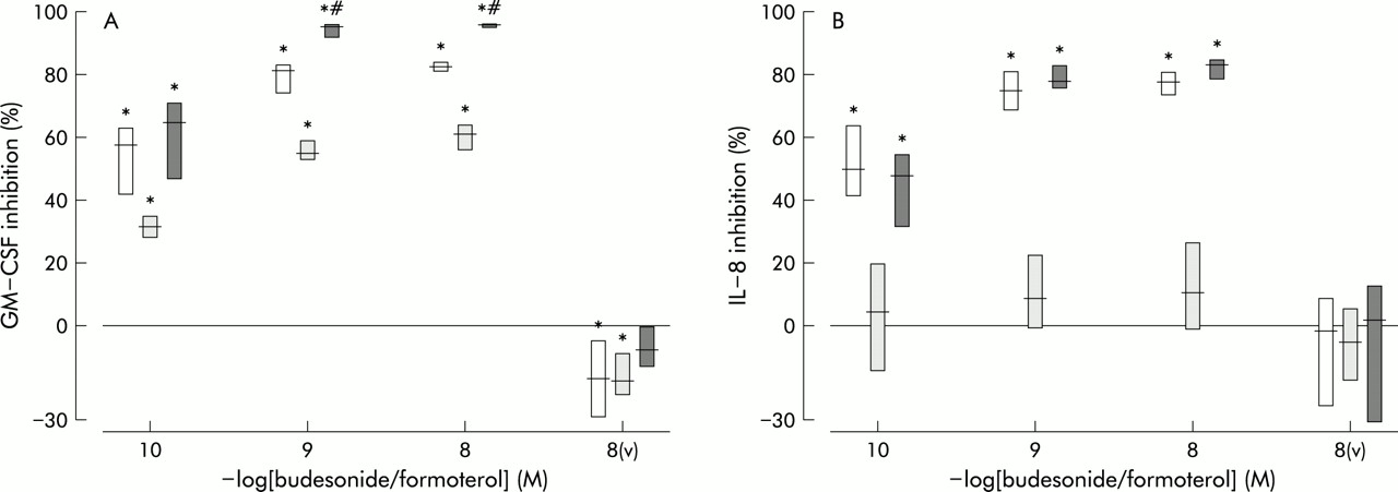
Human lung fibroblasts produced large quantities of the cytokine GM-CSF and the chemokine IL-8 after IL-1β stimulation (table 1). Budesonide and formoterol significantly inhibited IL-1β induced GM-CSF production in a dose dependent manner at all concentrations tested (fig 2A). The combination of budesonide and formoterol significantly inhibited GM-CSF production by 66 (47–71)% at 10−10 M, but this was not significantly different from the inhibition by budesonide alone (58 (42–63)%). At concentrations of 10−9 M and 10−8 M the inhibition by the combination of drugs was significantly larger than the inhibition induced by similar concentrations of the separate drugs, but not larger than the sum of inhibition. These effects were therefore defined as additive. Vehicle controls for budesonide and formoterol, but not the combination, significantly enhanced GM-CSF production by 120 (105–129)% and 120 (109–122)%, respectively. IC50 values for budesonide, formoterol, and the combination of the two drugs are presented in table 2.
Effects of budesonide (open bars), formoterol (dark shaded bars) or both (light shaded bars) in concentrations of 10−10–10−8 M on (A) GM-CSF and (B) IL-8 production of human lung fibroblasts induced by IL-1β (10 U/ml). Results are presented as median percentages of inhibition with interquartile range (n=6). v=vehicle control of 10−8 M budesonide, formoterol, or both. *p<0.05 compared with expression after IL-1β stimulation; #p<0.05 compared with inhibition of budesonide and formoterol alone (additive effect).
IL-8 production after IL-1β stimulation was significantly inhibited by budesonide in a dose dependent manner but, in contrast to the effect on GM-CSF, there was no additive effect of formoterol at the concentrations assessed (fig 2B). Moreover, formoterol alone did not significantly inhibit IL-8 production. Vehicle controls did not influence IL-8 production significantly.
Additional preincubation of either budesonide or formoterol separately 1 hour before the combination of the drugs was added showed a significantly stronger inhibitory effect on GM-CSF production than standard preincubation (not shown). This stronger inhibitory effect did not apply to IL-8 production.
DISCUSSION
This study shows that budesonide inhibits both GM-CSF and IL-8 production of human lung fibroblasts after IL-1β stimulation. Formoterol exerts an additive effect on the inhibition of IL-1β induced GM-CSF production, but not on inhibition of IL-8 production by budesonide. Formoterol also exerts mainly an additive effect on the inhibition of IL-1β induced ICAM-1 and VCAM-1 upregulation by budesonide.
Our observations suggest that the combination of budesonide and formoterol as therapeutic treatment may have an increased inhibitory effect on chronic inflammation in the airways of asthmatic individuals compared with the separate use of these drugs. Upregulation of adhesion molecules on resident pulmonary cells is diminished, which may lead to reduced infiltration of inflammatory cells. Production of GM-CSF is also diminished, which may lead to decreased activation and chemotaxis of eosinophils in the pulmonary tissue. Prevention of migration and activation of inflammatory cells probably results in better control of chronic and acute inflammation and less bronchoconstriction and hyperresponsiveness.
Our in vitro data are in accordance with data from clinical studies evaluating the effect of formoterol in addition to glucocorticoids. It has been shown that formoterol has an additive effect on inhaled glucocorticoids in reducing symptoms and the number of exacerbations and improving morning peak expiratory flow (PEF) and forced expiratory volume in 1 second (FEV1).1 Salmeterol and salbutamol alone did not inhibit IL-8 release from human airway smooth muscle cells but they enhanced the inhibition induced by dexamethasone and fluticasone.6 Salbutamol alone had an inhibitory effect on eotaxin production by human airway smooth muscle cells, and this effect was stronger when salbutamol was combined with dexamethasone or fluticasone.5 Results of an in vivo study in which reversion or prevention of formoterol induced β2 adrenoreceptor tolerance by systemic glucocorticoids was found also support the beneficial effects of combining glucocorticoids and β2 agonists.22 In contrast, there are also reports in which glucocorticoids do not prevent the development of tolerance induced by β2 agonists.23, 24
There are several reports of antagonistic actions of β2 agonists and glucocorticoids at the cellular level. Kankaanranta et al25 reported that, in contrast to glucocorticoids, β2 agonists delayed eosinophil apoptosis. The addition of salmeterol (long term exposure) to dexamethasone resulted in an antagonistic effect on the inhibition of superoxide production by eosinophils.26 In the same in vitro study an antagonistic effect of albuterol on dexamethasone induced eosinophil apoptosis was also found. On the other hand, albuterol did not antagonise the inhibition of GM-CSF and TNFα production of monocytes by budesonide27 and the long acting β2 agonist salmeterol had an additive effect on the inhibition of GM-CSF production in blood mononuclear cells by dexamethasone.15 Discrepancies in the abovementioned studies may be the result of differences in cell specificity, in the measured inflammatory variables, in the incubation time of the drug, or in the nature of the stimulus used.
Glucocorticoids act via AP-1 and NFκB by binding of GC-GR to these transcription factors.4 Through this mechanism they probably influence gene transcription of adhesion molecules28, 29 and cytokines.30. Formoterol stimulates β2 adrenoceptors which causes activation of the adenylyl cyclase system and a rise in intracellular cAMP levels, leading to PKA activation. PKA, in turn, is able to activate cAMP responsive element binding protein (CREB). CREB can interact with transcription factors such as AP-1 and NFκB, thus interfering with gene transcription.4
There are different possible interactions between glucocorticoids and β2 agonists ranging from synergistic or additional effects to antagonistic effects.4 The additive effects reported here probably result from the fact that both CREB and the GC-GR complex bind the transcription factors that are necessary for induction of gene transcription of ICAM-1, VCAM-1, and GM-CSF. An alternative possibility is that glucocorticoids increase transcription of the β2 adrenoceptor in lung tissue31 and prevent desensitisation of this receptor after long term exposure to β2 agonists.22
In contrast to GM-CSF production, IL-8 production is not inhibited by formoterol in the same experimental setting. However, IL-8 release from human smooth muscle cells is inhibited by salmeterol, but only when steroids are given concomitantly.6
We found similar additive effects when the sequence of formoterol and budesonide addition was varied. Only with GM-CSF production was the inhibition stronger when budesonide or formoterol were added before the combination of these drugs than without preincubation of the separate drugs. This can be explained by the longer total time of preincubation in the first two cases, and longer incubation times would probably remove this difference. The fact that this was not seen with ICAM-1 and VCAM-1 expression or IL-8 production may be because of the high sensitivity of GM-CSF production to the inhibitory effects of budesonide and formoterol. In contrast to our findings, Korn et al32 found an inhibitory effect of a β2 agonist on the activity of a glucocorticoid. The β2 agonist terbutaline inhibited the downregulation of α and β GR mRNA by budesonide in epithelial cells, but this effect was abrogated when budesonide was applied 4 hours before terbutaline. The authors concluded that, in order to avoid deleterious interference of β2 agonists with the activity of glucocorticoids, they should be administered in vivo a few hours after the glucocorticoid. However, a clinical study found no negative interaction when budesonide and terbutaline were used in combination.33 Our results are in agreement with the in vivo data.
In conclusion, formoterol had a mainly additive effect on the inhibition of ICAM-1 and VCAM-1 upregulation and GM-CSF production by budesonide in human lung fibroblasts. Inhibition of IL-8 production by budesonide was not affected by formoterol. Our in vitro data indicate that there is no reason, at the level of human lung fibroblast activation, why patients should not use these drugs simultaneously. As established in a clinical study,1 the beneficial effects may partially have been achieved through the additive inhibition of cellular infiltration resulting from diminished adhesion molecule expression and GM-CSF production of lung fibroblasts.
Acknowledgments
The authors thank Dr M Boorsma for critically reading the manuscript. This study was supported by a grant from AstraZeneca, The Netherlands.