Article Text

Abstract
Background: Despite having a work related deterioration in peak expiratory flow (PEF), many workers with occupational asthma show a low degree of within day diurnal variability atypical of non-occupational asthma. It was hypothesised that these workers would have a neutrophilic rather than an eosinophilic airway inflammatory response.
Methods: Thirty eight consecutive workers with occupational asthma induced by low molecular weight agents underwent sputum induction and assessment of airway physiology while still exposed at work.
Results: Only 14 (36.8%) of the 38 workers had sputum eosinophilia (>2.2%). Both eosinophilic and non-eosinophilic groups had sputum neutrophilia (mean (SD) 59.5 (19.6)% and 55.1 (18.8)%, respectively). The diurnal variation and magnitude of fall in PEF during work periods was not significantly different between workers with and without sputum eosinophilia. Those with eosinophilia had a lower forced expiratory volume in 1 second (FEV1; 61.4% v 83% predicted, mean difference 21.6, 95% confidence interval (CI) 9.2 to 34.1, p=0.001) and greater methacholine reactivity (geometric mean PD20 253 μg v 1401 μg, p=0.007). They also had greater bronchodilator reversibility (397 ml v 161 ml, mean difference 236, 95% CI of difference 84 to 389, p=0.003) which was unrelated to differences in baseline FEV1. The presence of sputum eosinophilia did not relate to the causative agent, duration of exposure, atopy, or lack of treatment.
Conclusions: Asthma caused by low molecular weight agents can be separated into eosinophilic and non-eosinophilic pathophysiological variants with the latter predominating. Both groups had evidence of sputum neutrophilia. Sputum eosinophilia was associated with more severe disease and greater bronchodilator reversibility but no difference in PEF response to work exposure.
- occupational asthma
- eosinophils
- neutrophils
- nitric oxide
- St George's Respiratory Questionnaire
Statistics from Altmetric.com
The normal and asthmatic cellular profiles of induced sputum are now well described1, 2 and sputum eosinophilia (eosinophils >2.2% non-squamous cells) occurs in most subjects with asthma.3 The cellular characteristics of induced sputum in occupational asthma are less well described and so far the findings in relation to exposure at work have been contradictory. Di Franco and co-workers reported significantly fewer sputum eosinophils and more neutrophils in occupational asthma due to low molecular weight agents than in non-occupational asthma.4 Lemiere et al found that subjects with occupational asthma had an increase in sputum eosinophils during periods at work compared with periods away from work and that this could be used to support the diagnosis.5 The diagnostic inclusion criteria for these studies were different and may explain some of the results; the former study required a diagnosis based on a positive specific inhalation challenge test, the latter required a fourfold change in non-specific bronchial reactivity. If, as some have suggested, there is a relationship between eosinophils and non-specific bronchial reactivity,2 the increase in eosinophils during workplace exposure in Lemiere's study may simply be reflecting the diagnostic inclusion criteria.
Serial measurement of peak expiratory flow (PEF) is the usual first line investigation of workers suspected of having occupational asthma. Although sensitivity and specificity have consistently been shown to be high,6–8 there is a degree of inter-observer and intra-observer variability in their interpretation. In particular, there is difficulty with those workers who show relatively small (<15%) but consistent falls in PEF during work periods and whose diurnal variability is considered to be within normal limits. While some would accept these as having occupational asthma if the clinical history was suggestive and they were exposed to a known sensitising agent, others might not. There are few available data regarding the airway inflammatory features associated with this sort of physiological response. We hypothesised that these workers would have a neutrophilic rather than an eosinophilic sputum inflammatory response. The aim of this study was to determine the sputum cellular profile of consecutive workers with occupational asthma induced by low molecular agents and to relate this to physiological measures of airway obstruction.
METHODS
Study protocol
Study subjects were recruited from sequential workers referred to the Occupational Lung Disease Unit at the Birmingham Chest Clinic who fulfilled our criteria for occupational asthma and who were exposed at work to a low molecular weight causative agent. In our clinic occupational asthma is diagnosed when a worker has wheeze/chest tightness and breathlessness temporally related to work exposure, with a latent interval between first exposure and first symptoms, and has at least one confirmatory test: serial measurement of PEF, specific bronchial provocation test, >3.2-fold change in non-specific bronchial reactivity in relation to workplace exposure, or specific IgE to a relevant low molecular weight agent. Measurement of specific IgE, assessment of non-specific bronchial reactivity within 24 hours of workplace exposure using the Yan technique9 (normal >2000 μg methacholine, equivalent to 10.22 μmol), and serial measurement of PEF are carried out at presentation. Specific bronchial provocation tests and repeat measurement of non-specific reactivity away from work are carried out later if indicated. The PEF record used for analysis in this study was the one immediately preceding sputum induction. If objective evidence supporting a diagnosis of occupational asthma was obtained, the subject was invited to attend for exhaled nitric oxide measurement and sputum induction. Sputum induction and measurement of exhaled nitric oxide was performed within 24 hours of a period of exposure to the causative agent at work of 3 or more consecutive days. Spirometric tests and bronchodilator reversibility were performed at this visit before sputum induction. Health related quality of life was assessed using the St George's Respiratory Questionnaire (SGRQ).10 Local ethical committee approval was obtained before the study and all subjects gave their consent to participate.
Subjects
Sixty one consecutive patients with confirmed occupational asthma who were still exposed to the causative low molecular weight agent at work were identified in an 18 month period. Forty five (73.8%) attended for sputum induction and 16 could not/did not wish to attend because of difficulty getting further time off work. An adequate sputum sample was obtained in 38 subjects (84.4%). Thirty six of these 38 subjects had serial PEF records diagnostic of occupational asthma (Oasys-2 score >2.511) and 12 also underwent specific bronchial challenge tests, all of which were positive. Specific IgE was not detected in the six subjects in whom a result is available. Two of the subjects had a history of pre-existing asthma. All subjects had a latent interval between first exposure and symptom onset (or deterioration in the cases with pre-existing asthma), suggesting that sensitisation had occurred in all cases. None of the subjects had clinical or radiographic evidence of interstitial lung disease.
Serial PEF records
Workers were requested to record PEF every waking 2 hours for 4 weeks including periods at and away from work. The best of three PEF readings were recorded on each occasion provided the best two readings were within 20 l/min of each other. Records were linearised,12 then plotted and analysed by Oasys-2. Oasys-2 is a computer program that uses discriminant analysis to compute a score between 1 and 4 indicating the likelihood of a PEF record showing occupational asthma. A score of >2.5 has a sensitivity of 75% and specificity of 94% for diagnosing occupational asthma.11 PEF records were also inspected visually by two experts for evidence of fabrication. Workers whose records were of inadequate quality—for example, containing fewer than four readings per day for the majority of the record—or of which there was doubt about the authenticity were excluded from entry into the study before sputum induction. PEF diurnal variability was calculated for days at work after days with less than four readings per day were excluded. The result was expressed as the mean of the maximum minus the minimum daily PEF divided by predicted PEF for each day. The absolute difference between the mean PEF on rest and work days and also the difference between the mean maximum rest day PEF and the mean minimum work day PEF (expressed as percentage maximum PEF) were calculated.
Exhaled breath nitric oxide
Exhaled breath nitric oxide levels (NO) were measured (Logan LR2000) during the plateau phase according to the ERS guidelines13 before sputum induction.
Induced sputum
Sputum induction was performed using a slightly modified version of the method described by Pavord et al.14 Lung function was measured before and every 5 minutes during the procedure. Salbutamol 200 μg was given via a metered dose inhaler before sputum induction. The subjects inhaled increasing concentrations of hypertonic saline (3%, 4%, and 5%) for 5 minutes at each concentration generated from an ultrasonic nebuliser (De Vilbiss Ultraneb 2000; output 6.2 ml/min, MMD 4.5 μm). If there was a 10–20% fall in forced expiratory volume in 1 second (FEV1) the concentration of hypertonic saline was not increased and if the fall in FEV1 was ≥20% the procedure was stopped altogether. Subjects were asked to blow their noses, rinse their mouths, and swallow the water before expectorating. Sputum plugs were separated from saliva. Selected portions were treated with 0.1% dithiothreitol (volume equal to four times the weight of selected sputum) for 15 minutes before being diluted with four volumes of Dulbecco's phosphate buffered saline (D-PBS). The suspension was filtered, then centrifuged at 2000 rpm for 10 minutes. The supernatant was removed and frozen and the cell pellet resuspended in D-PBS. Total cell count, cell viability, and squamous cell contamination was assessed using trypan blue in a Neubauer haematocytometer. Cytospin slides were obtained and stained with May Grunwald Giemsa. Differential counts were obtained from 400–600 non-squamous cells on two slides counted by a researcher who was unaware of the physiological response. Sputum eosinophilia was defined as eosinophils >2.2% of non-squamous cells based on 95% confidence limits of normal subjects from two large studies.1, 15
Repeatability of induced sputum
Sputum induction was repeated in 12 subjects following a 1 week period away from exposure at work.
Statistical analysis
Physiological and inflammatory marker data were normally distributed except for methacholine PD20, sputum eosinophil counts, and exhaled breath nitric oxide which were log transformed. A correction factor of 0.1 was added to eosinophil counts as some subjects had a count of 0%. Subjects who had a methacholine PD20 of >4800 μg were arbitrarily assigned a value of 9600 μg for analysis. Comparisons between eosinophilic and non-eosinophilic groups were performed using t tests. Latent interval and duration of symptomatic exposure were expressed as the median value and group comparisons were made using Mann-Whitney U tests. Differences in proportions were assessed using the Fisher's exact test. Pearson's correlation coefficient was used to test for linear relationships after log transformation where necessary. Multiple linear regression analysis was used to study models of physiological responses. All statistics were performed using SPSS for Windows 9.0.
RESULTS
The characteristics of the study subjects at the time of the investigation are shown in table 1. The median non-squamous cell counts (interquartile range) for all workers were: eosinophils 1% (0.2–4.3), neutrophils 61% (37.5–72.3), and macrophages 32.3% (23–44.9); cell viability was 77% (62.2–82) and squamous cell contamination was 6% (2.9–11.2). Only 14 of the 38 subjects (36.8%) had sputum eosinophilia. The frequency distribution of eosinophil counts is shown in fig 1. Differences between those with and without sputum eosinophilia are shown in table 1.
Characteristics of workers with occupational asthma with and without sputum eosinophilia

Frequency distribution chart of sputum eosinophil counts among workers with occupational asthma.
There was no difference in PEF diurnal variation or in the magnitude of the PEF response to work exposure in eosinophilic and non-eosinophilic subjects. Those with sputum eosinophilia had a lower prebronchodilator FEV1 (61.4% predicted v 83% predicted, mean difference 21.6, 95% CI of difference 9.2 to 34.1, p=0.001) and a lower FEV1/FVC (61.4% v 70.2%, mean difference 8.8, 95% CI 1.4 to 16.1, p=0.02). They had a worse health related quality of life (total SGRQ score 57 v 44.4, 95% CI of difference 2.5 to 22.7, p=0.016) with the symptom and impact components of the SGRQ being worse but not the activity component. Sputum eosinophilia was also associated with greater FEV1 reversibility to salbutamol (397 ml v 161 ml, mean difference 236 ml, 95% CI of difference 84 to 389, p=0.003) and a lower methacholine PD20 (geometric mean 253 μg v 1401 μg, p=0.007). Exhaled NO was significantly increased in eosinophilic subjects (geometric mean 10.4 ppb v 5.1 ppb, p<0.001); log NO correlated with log eosinophil count even when the non-eosinophilic subjects were considered separately (r=0.61, p<0.001 in all subjects and r=0.46, p=0.024 in non-eosinophilic subjects).
The presence of sputum eosinophilia was not related to atopy, duration of exposure, latent interval, or smoking history, nor did sputum eosinophilia relate to the causative agent (table 2). Non-eosinophilic subjects were receiving less treatment with inhaled steroids than eosinophilic subjects.
Causative agents
Bronchodilator reversibility was best expressed as the absolute value as there was less correlation with FEV1 % predicted than when expressed as % baseline (r=–0.28, p=0.088 for absolute value and r=–0.61, p<0.001 when expressed as % baseline; fig 2). Methacholine PD20 was also correlated with FEV1 % predicted (r=0.62, p<0.001; fig 3). Multiple linear regression analysis was used to determine whether methacholine reactivity or bronchodilator reversibility were related to eosinophilic inflammation independently of FEV1; 45% of the variation in log PD20 and 25% of the variation in bronchodilator reversibility could be explained by a model consisting of the independent variables FEV1 % predicted, log eosinophil count, and steroid treatment (table 3). The log eosinophil count showed no significant effect in the model for log PD20 (p=0.93) but did show a significant effect in the model for bronchodilator reversibility (p=0.045).
Multiple linear regression models of the form y = α + β1FEV1 + β2log(eosinophil) + β3inhaled steroids where (1) y = log PD20 and (2) y = bronchodilator reversibility (absolute value)
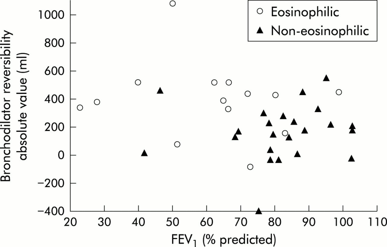
Bronchodilator reversibility for subjects with and without sputum eosinophilia as a function of FEV1 % predicted.

Methacholine PD20 for subjects with and without sputum eosinophilia as a function of FEV1 % predicted.
When eosinophilic and non-eosinophilic groups were considered separately, no significant correlation between log eosinophil count and bronchodilator reversibility was found within either group (r=0.19, p=0.39 in non-eosinophilic subjects, r=0.06, p=0.84 in eosinophilic subjects). This suggests that bronchodilator reversibility was related to the presence of sputum eosinophilia rather than the degree of eosinophilia.
The mean (SD) neutrophil count in those subjects who did not have sputum eosinophilia was 55.1 (18.8)% and 59.5 (19.6)% in those with eosinophilia. In non-eosinophilic subjects the neutrophil count was correlated with FEV1 % predicted (r=–0.46, p=0.022).
Of the 12 subjects in whom induction of sputum was repeated after a 1 week period away from work, six initially had sputum eosinophilia and six were non-eosinophilic. No subject changed to a different eosinophil status when induced sputum was repeated. There were no significant differences in eosinophil count between exposed and non-exposed subjects (median counts 2.2% v 2.5%, p=0.88). The intraclass correlation coefficient for eosinophil repeatability was 0.51.
DISCUSSION
We have shown that workers with occupational asthma caused by low molecular weight agents can be separated into eosinophilic and non-eosinophilic groups by induced sputum cell counts and that the non-eosinophilic group predominates. Repeatability of the presence or absence of sputum eosinophilia was shown in a proportion of subjects. The presence of sputum eosinophilia did not relate to atopy, causative agent, latent interval, or duration of symptomatic exposure. The eosinophilic group had a lower FEV1 that was not related to known confounding factors such as smoking history or treatment with inhaled steroids.
The findings of other studies of asthma in workers exposed to low molecular weight agents are somewhat contradictory. The median eosinophil count in our study was similar to that reported by Di Franco and co-workers who also reported that eosinophil counts in patients with asthma induced by low molecular weight agents were lower than in those with asthma due to high molecular weight agents or non-occupational asthma.4 Lemiere et al found sputum eosinophilia in nine out of 10 exposed workers.5 The different findings may in part reflect different diagnostic inclusion criteria. In Lemiere's study 80% of subjects had a fourfold or greater change in reactivity related to work exposure. These are likely to represent a subset of all occupational asthma as defined by specific bronchial provocation testing. Perrin et al showed that a greater than 3.2-fold change in responsiveness had a diagnostic sensitivity of 43% compared with specific challenge testing.6 It is possible that the control subjects in Lemiere's study may also have had occupational asthma but were not included as such because they did not fulfil the restrictive diagnostic criteria of the study. Our diagnostic inclusion criteria were less restrictive and made no prior assumptions as to the underlying mechanism. Obata et al, however, found an increase in sputum eosinophils during challenge with plicatic acid in nine consecutive subjects with western red cedar asthma.16 The same group reported that, of 21 consecutive exposed workers with western red cedar asthma, the mean eosinophil count of those on inhaled corticosteroids (n=16) was 1.7% compared with 3.7% in the five patients not on inhaled steroids17; it is unclear how many of these had sputum eosinophilia.
Although treatment with inhaled steroids tends to reduce sputum eosinophil numbers, there is evidence that low dose treatment might not do this significantly.18 Seventeen of the 24 subjects without sputum eosinophilia in our study were on no or low dose inhaled steroids, which suggests that treatment is unlikely to be a significant confounding factor.
Some authorities believe that PEF records showing diurnal variation within the normal range or those showing small changes in PEF (<15%) related to work exposure do not have occupational asthma.19 We originally hypothesised that those with low PEF diurnal variation had a different pathophysiology and would be non-eosinophilic. The results show no relationship between these PEF features and the presence or absence of sputum eosinophilia.
All subjects in our study had symptoms suggestive of asthma, and nearly all had a significant fall in PEF following exposure at work. There were 10 non-eosinophilic subjects who had normal methacholine reactivity and an FEV1/FVC ratio of >70%. Sceptics might argue that these subjects did not have asthma, were exaggerating their symptoms, and fabricated their PEF records to facilitate compensation. Three of these subjects underwent single blind bronchial provocation tests with nebulised solutions. All three had negative control challenges but had positive active challenges (two dual reactions and one late reaction). Six other positive specific challenge tests were in subjects who did not have sputum eosinophilia.
Several studies of airway inflammation in non-occupational asthmatic subjects have reported heterogeneity of cell counts. Wenzel et al reported two pathophysiological subtypes amongst severe asthmatics, one without evidence of eosinophil infiltration in bronchial biopsy specimens.20 Gibson et al found most of the asthmatic subjects in their study to be non-eosinophilic with an increased neutrophil count.21 The role of the neutrophil in asthma is unclear. Increased numbers of airway neutrophils have been reported in severe asthmatics22 and the presence of increased numbers of neutrophils has been demonstrated in bronchial biopsy specimens from non-atopic asthmatics23 and workers with grain induced occupational asthma.24 Even subjects without sputum eosinophilia in our study had evidence of a neutrophilic bronchial inflammatory response unrelated to smoking history. Their sputum neutrophil count was significantly raised (mean neutrophil count 55.1%, 95% CI 47.6 to 62.6) compared with that previously reported in normal individuals. Spanevello et al found a mean neutrophil count of 27.3% (95% CI 24.7 to 29.9) in 96 normal subjects1 and Belda and colleagues reported a mean of 37.5% (CI 33.5 to 41.5).15 We found a significant relationship between sputum neutrophils and FEV1 % predicted in non-eosinophilic subjects.
There will inevitably be some selection bias of the workers studied, as with most studies in occupational and non-occupational asthma. Workers with a diagnosis of occupational asthma who had been removed from exposure by the time they were seen may have had more severe disease but were not included in this study. We tried to minimise selection bias by trying to recruit consecutive exposed subjects with a diagnosis confirmed by any of the accepted objective methods.
Multiple linear regression models were used to investigate whether the differences in bronchodilator reversibility and methacholine reactivity observed between eosinophilic and non-eosinophilic groups were independent of FEV1. The non-eosinophilic group had a significantly lower bronchodilator response than the eosinophilic group that was independent of FEV1. However, differences between groups in methacholine reactivity were not independent of FEV1.
The different pathophysiology of airway disease of workers with asthma induced by low molecular weight agents without sputum eosinophilia suggests a different disease process than that which occurs in eosinophilic asthma. The lack of bronchodilator reversibility suggests less bronchial smooth muscle involvement, which might occur if there was less distal airway disease or if mucosal oedema predominated. Some physicians might feel that some of these workers do not fulfil their definition of asthma. Differences in opinion may be due to the imprecise definition of asthma. Significant differences in mean PEF at rest and at work are unlikely to occur in normal individuals, although it is possible that “irritant” responses could cause a deterioration in PEF during work periods. Differentiating between irritants and sensitisers is not always easy as specific IgE is usually not detectable in asthma induced by low molecular weight agents. In practice the distinction is made on the basis of the history—for example, the presence of a recognised sensitiser in the workplace, a period of latency between first exposure and symptoms indicating some form of sensitisation. The disease in the non-eosinophilic subjects resembles asthma more than any other condition and cannot be differentiated on the basis of clinical history or PEF responses. Most do not expectorate sputum on a daily basis, hence occupational bronchitis would seem an inappropriate label. Many of these workers have a significant impairment of health related quality of life. We feel that it would still seem appropriate to label these workers as having asthma as they have evidence of airflow obstruction reversible over short periods of time in relation to specific exposure at work. It might be appropriate to differentiate them further by describing them as having occupational non-eosinophilic or neutrophilic asthma. Further studies into the nature of airway inflammation through bronchial biopsies are warranted as well as studies into the prognosis in these workers.
In conclusion, asthma caused by low molecular weight agents can be separated into eosinophilic and non-eosinophilic pathophysiological variants with the latter predominating. Both groups had evidence of sputum neutrophilia. Sputum eosinophilia was associated with more severe disease and greater bronchodilator reversibility, but no difference in peak expiratory flow response to work exposure.
Acknowledgments
The authors would like to thank all members of the Oasys team for their help and Dr Shakir Hussain for statistical advice. The project is funded by the European Chemical Industry Council (CEFIC). Dr Anees is also supported by a grant from the COLT foundation.