Article Text

Abstract
BACKGROUND Theophylline is widely used in the treatment of asthma, and there is evidence that theophylline has anti-inflammatory or immunomodulatory effects. A study was undertaken to determine whether theophylline added to low dose inhaled steroids would be as efficacious as high dose inhaled steroids in asthma.
METHODS In a study in general practice of 155 recruited asthmatic patients with continuing symptomatic asthma while on 400 μg beclomethasone dipropionate (BDP) daily and inhaled β2 agonist as required, the effect of (1) continuing low dose inhaled steroids alone (LDS, 200 μg BDP twice daily), (2) low dose inhaled steroids plus low dose theophylline (LDT, 400 mg daily), or (3) high dose inhaled steroids (HDS, 500 μg BDP) over a six month period was examined.
RESULTS One hundred and thirty patients completed the study. Between group comparison using analysis of variance showed no overall differences in peak flow measurements, diurnal variation, and symptom scores. Changes in evening peak flows approached significance at the 5% level (p=0.077). The mean improvement in evening peak flow in the LDT compared with the LDS group was 20.6 l/min (95% confidence interval (CI) –2.5 to 38.8). In the LDT group there was an increase in evening peak flows at the end of the study compared with entry values (22.5 l/min), while in the LDS and HDS groups evening peak flows increased by 1.9 and 8.3 l/min, respectively. There was no significant difference in exacerbations or in side effects.
CONCLUSION There were no overall significant differences between the low dose steroid, low dose steroid with theophylline, and the high dose steroid groups. The greatest within-group improvement in evening peak flows was found after theophylline. A larger study may be necessary to show significant effects.
- asthma
- theophylline
- inhaled corticosteroids
- dosage
- general practice
Statistics from Altmetric.com
Theophylline is used worldwide for the treatment of asthma and is usually prescribed as bronchodilator therapy, although its bronchodilator efficacy is lower than that of β2adrenergic agonists. Recent studies indicate that theophylline also has anti-inflammatory effects, as shown by its inhibition of the late response to allergen, together with a reduction in bronchial mucosal eosinophils induced by allergen challenge in patients with asthma.1 Theophylline at serum levels below the accepted therapeutic range (<10 mg/l) provided improvements in lung function in patients with moderately severe asthma already established on high dose inhaled steroids, together with a reduction in mucosal CD4+ and CD8+ T cells.2 The clinical usefulness of theophylline has been extended by the recent finding that the addition of theophylline to a medium dose of inhaled steroids (for example, budesonide 800 μg/day) was more effective in the control of asthma than a doubling of the dose of steroid (for example, budesonide 1600 μg/day).3 Similar benefits were also obtained when theophylline was added to low dose inhaled steroids (beclomethasone dipropionate (BDP) 400 μg/day) rather than using a higher dose of inhaled steroids alone (BDP 800 μg/day).4
We have re-investigated the potential clinical benefits of adding low dose theophylline to low dose inhaled corticosteroids in a larger population of patients with asthma over a longer period of time (six months) in a general practice setting. This study was particularly designed to see whether the benefit of adding low dose theophylline was sustained over time, and whether this was superior or equivalent to doubling the dose of inhaled steroids. Furthermore, in contrast to previous studies we have included a group of control patients who remained symptomatic on low dose inhaled corticosteroids, in an attempt to assess the real benefits of adding low dose theophylline. This was a double blind multicentre trial in general practice involving 155 recruited patients.
Methods
PATIENTS
Two hundred and forty nine non-smoking adult asthmatic adults aged between 18 and 65 who met the diagnostic criteria of the American Thoracic Society for asthma were recruited for the study5from participating general practice units in the UK. The patients had persistent asthmatic symptoms in the six weeks before the study while being treated with low dose inhaled steroids (200 μg BDP, 100 μg fluticasone propionate, or 200 μg budesonide twice daily), with or without inhaled short acting β2 agonists as required (⩽14 inhalations/day). They were also required to have a baseline peak expiratory flow rate (PEF) ⩾50% of the predicted normal, with at least 15% variability in PEF (amplitude % max) during the run in period, and asthmatic symptoms on at least three of the last seven days of the run in period. All randomised patients had no contraindication for the use of theophyllines and had not experienced an exacerbation of their asthma during the six weeks before the study. Patients taking long acting inhaled or oral β2 agonists or any other asthma therapy were excluded. All patients gave their informed consent and the study was approved by the local ethics committees.
STUDY DESIGN
The study was a randomised, double blind, parallel group study and was controlled with a group of patients treated with low dose steroids alone. The treatment regimens consisted of two study periods designed to compare the effects of the following three treatments: (1) low dose steroids consisting of 200 μg inhaled BDP twice daily plus placebo tablets (LDS); (2) low dose steroids (200 μg inhaled BDP twice daily) plus low dose theophylline, consisting of slow release theophylline tablets 200 mg (Phyllocontin slow release) twice daily (LDT); and (3) high dose steroids (500 μg inhaled BDP twice daily plus placebo tablets (HDS)). The treatments were randomised in balanced complete blocks with a block size of 3 and complete blocks of patient supplies were allocated to study centres.
The study periods included a run in period of two weeks when patients were screened at visit 1, issued with symptom and peak flow diary cards, together with a peak flow meter (Clement Clarke, Harlow, Essex UK). They then entered the one week run in period during which they received inhaled BDP 200 μg twice daily and were asked to chart their asthma symptoms and morning and evening PEF. They re-attended at the end of the run in period (visit 2) when their condition was re-assessed and fulfilment of the entry criteria was re-confirmed. Patients who satisfied these criteria entered the treatment period and were randomly allocated to one of three treatments. During the treatment period patients attended visits at monthly intervals for continual assessment (visits 3–8) and continued to monitor their symptom scores and PEF daily. All patients were asked to complete a Juniper asthma quality of life questionnaire6 at the start and end of the treatment periods. The primary outcome measures were mean morning and evening PEF as an assessment of clinical benefit from treatment. The secondary variables assessed were diurnal variation of PEF, use of short acting β2 agonists, symptom scores, asthma exacerbation counts, and quality of life.6
STATISTICAL ANALYSES
All efficacy data were summarised by treatment group as mean (SD) values. Diary card data were summarised for the eight days before each visit, for the run in period (run in), and for the last seven days of the treatment period (end). The changes between run in and end were calculated and compared between groups using analysis of variance (ANOVA). Data were also analysed within treatment groups using a pairedt test when a p value of <0.1 was achieved. The diurnal variability for each 24 hour interval was calculated by using the amplitude % maximum defined as the highest PEF minus the lowest PEF divided by the highest PEF. The mean values for the last seven days of the run in period and for the end of the assessment period were used to assess the effects of the three different treatments. The data from the quality of life questionnaire were summarised by each of the domains including activity, symptoms, emotions, and environment.
We have estimated that, with 50 completing patients in each treatment group, the study would have an 80% power at the 5% significance level to detect a difference between treatments in the change in morning PEFR from baseline of 35 l/min.
Results
Of the 249 patients recruited, 94 patients failed the run in and 155 patients fulfilled the inclusion criteria, were randomised for treatment, and completed the study. Fifty two received high dose steroids (HDS), 49 received low dose steroids and low dose theophylline (LDT), and 54 patients received low dose steroids (LDS). Their demographic characteristics and baseline data from the end of the run-in period are presented in table 1. The three groups were not statistically different with respect to demographic and baseline lung function data.
Mean (SD) patient demographic and baseline data at end of run in period
Of the 155 randomised patients, 25 patients withdrew (five from the HDS group, 11 from the LDT group, and nine from the LDS group). The reasons for withdrawal are shown in table 2.
Reasons for withdrawal from study
HOME RECORDINGS OF PEF AND PEF VARIABILITY
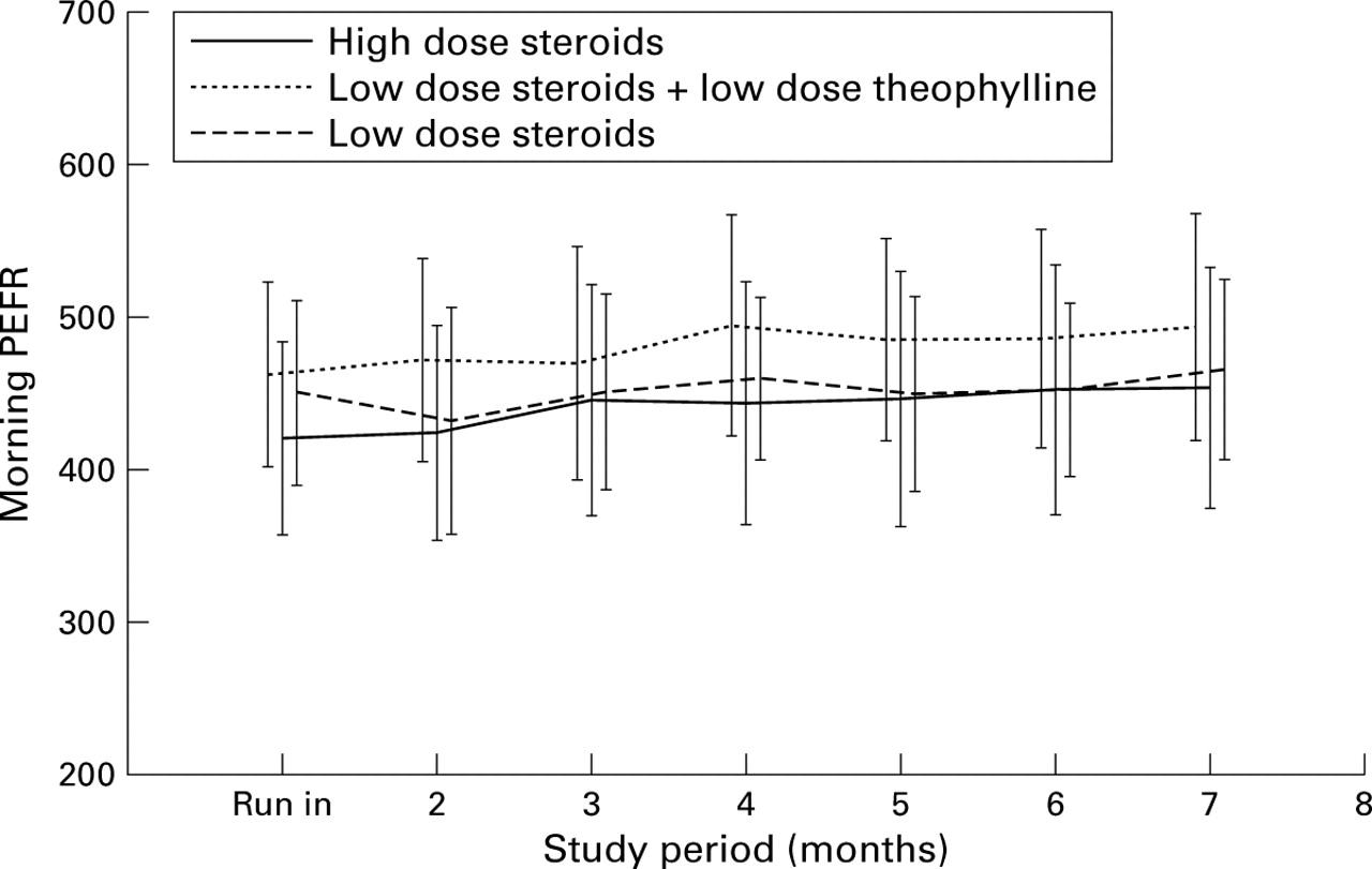
Analysis of the change in morning and evening peak flow showed no significant differences between the three groups (table 3). Between group ANOVA showed a trend towards significance for the evening PEF (p=0.077). Within group comparison showed that there was a significant improvement in mean morning PEF in patients taking HDS (from 395.8 (96.7) to 415.3 (112.7) l/min, p=0.007) and LDT (from 427.7 (85.0) to 449.5 (100.6) l/min, p=0.006; fig 1). Patients in the LDT group had a significant improvement in evening PEF from baseline (from 446.5 (86.5) to 469.1 (101.2) l/min, p=0.002; fig 2) but not those in the HDS or LDS groups. These improvements were maintained during the six month study period. There was no significant difference in diurnal variation between the groups, but within groups all three treatments led to small reductions in PEF variability.
Mean (SD) peak expiratory flow (PEF), β agonist usage, and symptom scores at end of study

Mean (SD) morning peak expiratory flow rate (PEFR) during the six month study for the three parallel groups of patients on low dose steroids, low dose steroids plus low dose theophylline, and high dose steroids. PEFR improved significantly during high dose steroid and low dose steroid plus theophylline (p=0.006).

Mean (SD) evening peak expiratory flow rate (PEFR) during the six month study for the three parallel groups of patients on low dose steroids, low dose steroids plus low dose theophylline, and high dose steroids. PEFR improved significantly during low dose steroid plus theophylline (p=0.002).
USE OF SHORT ACTING β2 AGONISTS AND SYMPTOM SCORES
There was no significant change in the use of short acting β2 agonists or asthma symptoms between the three groups, but all three groups improved from baseline with respect to medication usage and morning asthma symptom scores (table 3).
EXACERBATIONS
During the six month study period 20 patients required prednisolone for moderate symptomatic exacerbation of asthma. Eight patients (5.2%) taking HDS, three (1.9%) taking LDT, and 11 (7.1%) taking LDS reported exacerbations. These differences were not statistically significant.
QUALITY OF LIFE QUESTIONNAIRE
There were improvements in all four domains; however, there were no significant differences in the responses between the three groups (table 4).
Quality of life assessment and change in quality of life
SIDE EFFECTS
The side effects encountered during the study are summarised in table 5. There were no significant differences between the treatment groups for any of the commonly reported symptoms.
Volunteered side effects
Discussion
We found that there was a trend for improved evening PEF when theophylline was added to low dose inhaled steroids in patients with uncontrolled asthma. Within group analysis showed that the evening PEF did not change significantly in the low dose or high dose steroid groups. There were also some improvements in morning PEF in patients who received either low dose theophylline or high dose steroids. However, overall, there were no significant differences between the groups for the changes in morning PEF, diurnal variation, symptom scores, and quality of life measures. Our data indicate that, in the primary care setting, the addition of low dose theophylline to low dose steroids in patients not well controlled on the latter may be beneficial in improving evening peak flows. It is not possible to conclude from the current study whether the effect of adding theophylline in this setting is equivalent or better in efficacy than increasing the dose of steroid. Nevertheless, our data are in accordance with earlier studies3 ,4 which showed that theophylline provides clinical benefit under these circumstances, but the present study indicates that this can also be done in the primary care setting.
Previous studies addressing the issue of combination treatment with oral theophylline and inhaled corticosteroids have drawn criticism because of relatively small numbers, the lack of a control group, and the short duration of the study (six weeks to three months).3 ,4 We have addressed these issues in our study and have made power calculations which indicated the need for at least 50 patients in each of the three arms of the study. Because of a number of patient withdrawals during the study, we had less than 50 patients in each group with 47, 38, and 45 in the high dose, theophylline, and low dose groups, respectively. This could be one reason why statistical significance was not achieved. Other possibilities include the carryover effect of improved compliance with inhaled steroids, particularly in the low dose steroid group, since patients are more likely to be compliant when closely observed in a clinical trial. Evidence for improved lung function occurred during the run in period since many patients could not be enrolled as their symptoms disappeared and peak flows improved during the six week run in period.
The obvious comparison of these clinical end points in our study is with those in the study by Greening et al 7 in which the effect of adding salmeterol to inhaled low dose inhaled steroids was compared with high dose inhaled steroid treatment.7 A significant improvement was found in morning and evening PEF (28 l/min and 19 l/min, respectively) from baseline and significant differences in favour of salmeterol in symptoms, medication usage, and diurnal variation compared with high dose steroids. There was, however, no difference in exacerbations although a study by Pauwels et al 8 designed to examine this specific point showed a reduction in exacerbations. The results of our study are comparable with those of the salmeterol study in that we found similar improvements in PEF, PEF variability, and symptoms within the treatment groups, and no significant difference between the groups at the end of the study. We also found no statistical difference in the number of exacerbations between the groups, although there was a trend towards fewer exacerbations with low dose theophylline. The mean changes in peak flows after the addition of theophylline were therefore comparable to those after addition of long acting β2 agonists, indicating that these effects are clinically significant.
Although the mode of action and anti-inflammatory activity of theophylline continues to be debated, the clinical usefulness of this treatment for asthma is further supported by the results of the present study. Theophylline fulfils many aspects of the needs in the treatment of asthma. It is a bronchodilator and may exert some anti-inflammatory effects, and it exhibits inhibitory effects on many cell types. For example, theophylline inhibits the degranulation and release of mediators including platelet activating factor, leukotriene C4 and basic proteins9 ,10 from eosinophils. It inhibits proliferation and activation of T lymphocytes.11 Withdrawal of theophylline treatment leads to an increase in T cells within the airway mucosa of patients with asthma.2 Theophylline also attenuated the generation of pro-inflammatory cytokines such as IL-512 whilst also upregulating the expression of the anti-inflammatory cytokine IL-10.13 The improvement in PEF in our patients was sustained throughout the study, which suggests the absence of tachyphylaxis, which has also been reported with long acting β2 agonists.14
Recent studies of compliance with treatment in asthma have shown that adherence to inhaled prophylactic treatment is generally poor.15 Comparative studies have suggested better compliance with theophylline than with inhaled steroids.16Another potentially important consideration is the evaluation of the cost of treatment strategies. Theophylline is cheaper than either inhaled steroids or long acting β2 agonists. Clearly, a more comprehensive pharmacoeconomic evaluation of all potential factors is required.
Although this study did not specifically assess the anti-inflammatory activity of theophylline, it is probable that immunomodulation could account for some of the observations since we used a dose of theophylline that achieved concentrations below the therapeutic bronchodilator range of 10–20 mg/l. Although we did not measure serum theophylline levels in these patients, previous studies have shown that levels of 8.7 mg/l were achieved in patients treated with slow release theophylline in a dose of 500–750 mg/day.3 Moreover, the access of general practitioners to clinical laboratories and the inconvenience of measuring serum theophylline levels are important considerations in community practice, hence the choice of a dose of theophylline that would provide “safe” levels below 10 mg/l.
Our study has shown that the addition of low dose theophylline may be beneficial in patients whose asthma is not optimally controlled on low dose steroid. Although the role of inhaled steroids in the management of asthma is not in question, alternative strategies aimed at achieving control of asthma symptoms with low dose inhaled steroids may be safer, more attractive, and cheaper with the addition of low dose theophylline than using higher doses of inhaled steroids. Our study gives some support to the option of adding low dose theophylline to low dose inhaled steroids, an option that can be readily implemented in general practice. The benefits are at least comparable to the use of long acting β2 agonists at this step, but long acting β2 agonists are not likely to possess anti-inflammatory activity and are more expensive. However, a larger study with greater power is necessary to confirm these findings.
Acknowledgments
This study was supported by Napp Laboratories Ltd.