Article Text

Download PDFPDF

A large chest wall tumour in an asymptomatic 15-year-old girl
Free
  1. K-C Chen1,
  2. W-C Lin2,
  3. Y-C Lee1,3,
  4. J-S Chen1,
  5. H-H Hsu1
  1. 1
    Department of Surgery, National Taiwan University Hospital and National Taiwan University College of Medicine, Taipei, Taiwan
  2. 2
    Department of Pathology, National Taiwan University Hospital and National Taiwan University College of Medicine, Taipei, Taiwan
  3. 3
    Department of Traumatology, National Taiwan University Hospital and National Taiwan University College of Medicine, Taipei, Taiwan
  1. Dr H-H Hsu, Department of Surgery, National Taiwan University Hospital, No 7, Chung-Shan South Road, Taipei, Taiwan; ntuhfred{at}yahoo.com.tw

Statistics from Altmetric.com

Request Permissions

If you wish to reuse any or all of this article please use the link below which will take you to the Copyright Clearance Center’s RightsLink service. You will be able to get a quick price and instant permission to reuse the content in many different ways.

CLINICAL PRESENTATION

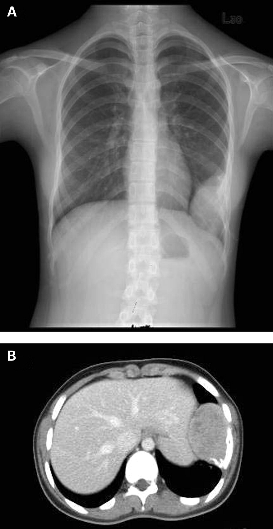
A 15-year-old girl that had plain chest x ray as part of a routine medical examination was found to have a large left lower chest wall mass associated with rib destruction that was later shown to be painless and non-palpable (fig 1A). Chest CT revealed a 7×11 cm pleura based mass located in the left lower thorax and featured multiple rib destruction and downward depression of the left hemidiaphragm (fig 1B).

Figure 1 (A) Chest plain film revealing a large, well defined left chest wall tumour with obvious rib destruction. (B) Thoracic CT revealing a large expansile tumour associated with osteolytic and sclerotic changes to a number of the patient’s ribs.

CT guided biopsy revealed a lesion with clusters of melanin–laden tumour cells. These tumour cells were positive for HMB-45 and S-100 protein immunohistochemically. The cytoplasmic pigments could be bleached by superoxidative agent and were positive by Fontana Masson stain, consistent with melanin pigments in nature. The skin overlying the tumour was intact, and the tumour was impalpable. Thorough skin examination over her whole body by an experienced dermatologist did not reveal any suspicious skin lesion. No other abnormality was demonstrated on whole body image studies.

QUESTION

What is the likely diagnosis and how should this be confirmed?

See page doi: 10.1136/thx.2007.087982 for answer

This case is submitted by:

View Abstract

Footnotes

  • Competing interests: None.

Linked Articles

  • Miscellaneous
    BMJ Publishing Group Ltd and British Thoracic Society