Article Text

Abstract
Background: A study was undertaken to analyse the potential prognostic value of the immunohistochemical expression of cyclooxygenase-2 (COX-2) and p27 in 29 malignant mesotheliomas already screened for the expression of p21 and p53.
Methods: Immunohistochemistry was used to determine the expression of COX-2 and p27. The correlation with survival of these factors and of p21 and p53 expression was assessed by univariate and multivariate analyses.
Results: A positive statistically significant correlation was found between p27 and p21 expression (p<0.0001), but there was a negative correlation between COX-2 expression and both p27 (p = 0.001) and p21 (p<0.0001). No statistically significant correlation was recorded between p53 and all the other immunohistochemical parameters. Univariate analysis showed that overall survival was strongly influenced by p21, p27, and COX-2 expression, but multivariate Cox regression analysis showed that the only immunohistochemical parameter to influence overall survival of patients with mesothelioma was COX-2.
Conclusions: These findings suggest that COX-2 expression may be a useful prognostic parameter for mesothelioma.
- mesothelioma
- cyclooxygenase-2
- cell cycle inhibitors
- prognosis
- survival
Statistics from Altmetric.com
Malignant mesothelioma is a rare, highly aggressive tumour which accounts for less than 1% of all cancer deaths in the world.1 This neoplasm arises from the surface serosal cells of the pleural (>90% of cases), peritoneal, and pericardial cavities and from the tunica vaginalis of the testis.2 Although the association between exposure to asbestos and the development of mesothelioma is commonly accepted, the exact mechanism whereby asbestos induces the mesothelioma is unknown.3 Several studies report the presence in some mesotheliomas of DNA encoding SV40 T antigen or SV40 T antigen protein expression, suggesting that the presence of this viral gene may also be associated with the pathogenesis of this neoplasm.4 The prognosis is generally poor with a reported median survival of 4–12 months in either untreated or treated (surgery, radiotherapy, or chemotherapy) patients.5 Moreover, mesothelioma has proved resistant to classical chemotherapeutic and radiation regimes and the natural history has not been influenced by standard therapy.6 Several clinical prognostic factors have been tentatively correlated to survival, histological type and performance status being the most valuable.5,7–9
Cyclooxygenase-2 (COX-2) has been implicated in carcinogenesis of several neoplasms through the downregulation of cell mediated immunity, promotion of angiogenesis, inhibition of apoptosis, and the formation of carcinogenic metabolites such as malondialdehyde.10–12 Furthermore, COX-2 overexpression has been noted in many solid tumours and has been correlated with a worse prognosis in colorectal cancer, non-small cell lung cancer, and gastric cancer.13–15 It has been shown that cultured human mesothelial cells contain cyclooxygenase activity16 and that COX-2 expressing mesothelioma cell lines are associated with increased proliferative and invasive potential.17 A recent study showed that COX-2 expression is a strong prognostic factor in human mesothelioma, which contributes independently to the other clinical and histopathological factors in determining a short survival.18 It has been proposed that COX-2 exerts its influence on mesangial cell proliferation in vitro by a novel mechanism involving the tumour suppressor p53 and the cell cycle inhibitors p21 and p27,19 and several recent studies have investigated the potential prognostic value of p53, p21 and p27 in malignant mesotheliomas.20–24
A study was undertaken to analyse the potential prognostic value of the immunohistochemical expression of COX-2 and p27 in 29 malignant mesotheliomas already screened for the expression of p21 and p5323,24 and for the presence of SV40-like sequences.25 The correlation with survival of each factor in univariate and multivariate analysis was assessed.
METHODS
Clinical data and tumour specimen acquisition
All patients were treated at the Second University of Naples between 1980 and 1996. Clinical data were obtained by retrospective chart review. Survival was determined from the date of initial surgery. Follow up was available for all patients. No patients were excluded from survival analysis because of death unrelated to cancer, and no patients were lost during follow up. The two patients not included in the analysis of survival were excluded because they died in the postoperative period from surgery related complications.
All patients underwent cytoreductive surgery and 13 patients were further treated with radiotherapy or chemotherapy. Only in three cases did the clinical history show a clear exposure to asbestos, but this does not mean that the other patients were not also exposed to asbestos. In fact, all the patients were living in Campania, a region where contamination with asbestos is very high. Moreover, cases were selected who were still available for immunohistochemical studies from the original group of 35 already characterised for the presence of SV40 sequences.25
Tissue from 29 malignant mesothelioma specimens (16 epithelioid, six sarcomatoid, and seven mixed mesotheliomas) obtained from open biopsies or pleurectomies were collected and fixed in 10% formalin before being embedded in paraffin.
Histological examination
Five μm sections were cut from the formalin fixed, paraffin embedded samples and stained with haematoxylin and eosin. The histological diagnosis was re-examined by two pathologists (AB and FB) according to the World Health Organization histological typing of tumours. In addition, the most representative blocks were selected to be cut into new 5 μm thick sections for immunohistochemical studies.
Immunohistochemistry
All 29 cases had been assessed by immunohistochemistry for the presence of COX-2 and p27. Five μm sections were cut from each specimen, mounted on glass, and dried overnight at 37°C. All sections were then deparaffinised in xylene, rehydrated through a graded alcohol series, and washed in phosphate buffered saline (PBS). PBS was used for all subsequent washes and for antibody dilution. Endogenous peroxidase activity was blocked by 5% hydrogen peroxide. The primary antibodies for p27 (mouse monoclonal; sc-1641, Santa Cruz Biotechnology, CA, USA) and for COX-2 (goat polyclonal; sc-1745, Santa Cruz Biotechnology) were applied at room temperature for 1 hour at a dilution of 1:100. The optimal working dilution was defined on the basis of titration experiments. The sections were then immunostained with the streptavidin-biotin system (Dako, Carpintera, CA, USA) using diaminobenzidine (DAB) as the final chromogen and haematoxylin as the nuclear counterstain. Negative controls for each tissue section were prepared by omitting the primary antibodies. A suitable positive control was run with each set of slides. All samples were processed under the same conditions.
The experimental conditions for the immunohistochemical analysis of p21 and p53, as well as for the detection of SV40 sequences, have been described elsewhere.23–25
Scoring and quantification of immunoreactivity
Two observers (AB and SS) estimated the staining pattern of p27 and COX-2 separately. For p27, each specimen was scored for the percentage of positive nuclei (<10% and >10% of cells expressing p27). For COX-2, staining intensity was scored as 0 (negative), 1 (weak), 2 (medium), and 3 (strong). The extent of staining was scored as 0 (0%), 1 (1–25%), 2 (26–50%), 3 (51–75%), and 4 (76–100%) according to the percentage of positive stained cells. The sum of the intensity and extent scores was used as the final staining score (0–7). The level of concordance, expressed as the percentage of agreement between the observers, was 93% (27 of 29 specimens). In the remaining specimens the score was obtained after collegial revision and agreement. The scoring and quantitation of immunoreactivity for p21 and p53 has been described elsewhere.23,24
To select the cut off point for COX-2 expression the samples were divided into two groups (0–4 and >4) to give a low expression and high expression group. For all the other markers a cut off point of 10% was chosen because they are all nuclear molecules involved in cell cycle regulation whose loss of expression is strictly connected with tumour progression. A value of 10% therefore indicates whether or not the samples are expressing each of these markers.
Statistical analysis
Fisher’s exact test was used to assess the relationship between ordinal data (correlation matrix between immunostaining parameters). The correlation between COX-2, p27, and p21 was performed on the original ordinal units using the Spearman test. Univariate survival analysis for each prognostic variable on overall survival was estimated according to the Kaplan-Meier method.26 The terminal event was death attributable to cancer. The statistical significance of the differences in survival distribution between the prognostic groups was evaluated by the log-rank test.27 The Cox proportional hazards model was applied to the multivariate survival analysis.28 The prognostic variables on overall survival included histological types, COX-2, p21, p27, and p53 expression. p values of <0.05 were regarded as statistically significant in two tailed tests. SPSS software version 10.00 (SPSS, Chicago, USA) was used for statistical analysis.
RESULTS
Histology and immunohistochemistry
The results of immunohistochemical analysis of 29 mesothelioma specimens are shown in table 1. Histologically, there were 16 epithelioid, six sarcomatoid, and seven mixed mesotheliomas. COX-2 staining was always cytoplasmic, while p53, p21 and p27 staining was preferentially nuclear although cytoplasmic positivity was occasionally seen for p21 and p27. Both cytoplasmic and nuclear stainings were considered positive. Figure 1 shows some typical immunohistochemical stainings for COX-2 (fig 1A and B) and p27 (fig 1C and D). p53 and p21 immunostainings on the same group of mesothelioma samples have been reported elsewhere.23,24
Characteristics of study patients
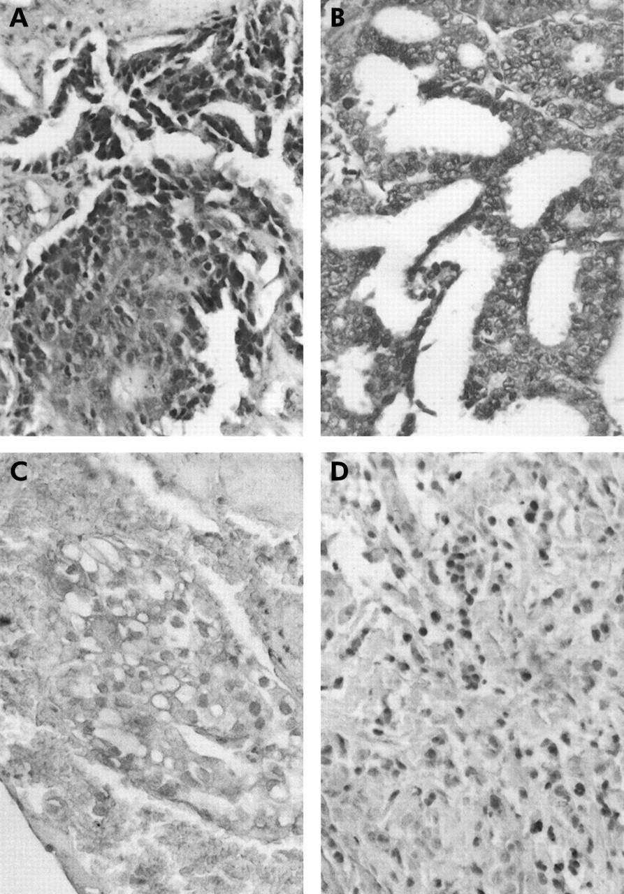
Immunhistochemical stainings of COX-2 (A and B) and p27 (C and D) in human pleural mesotheliomas (original magnification ×250).
Correlation between immunohistochemical and pathological parameters
A positive statistically significant correlation was recorded by rank correlation matrix between p27 and p21 expression, and a negative correlation was found between COX-2 expression and both p27 and p21 (table 2). Interestingly, there was no statistically significant correlation between p53 and all the other immunohistochemical parameters. No correlation was seen between immunohistochemical parameters and mesothelioma histological type.
Rank correlation matrix (and statistical significance) between molecular markers in patients with malignant mesothelioma
Immunohistochemical and pathological parameters and patient survival
Univariate analysis showed that overall survival was strongly influenced by p21, p27, and COX-2 expression. The median survival in patients with low p21 or p27 expression was longer than in those with high p21 or p27 expression. The median survival in patients with low COX-2 expression was 14 months compared with 5 months in those with high COX-2 expression. On the other hand, p53 expression and histological type did not influence the overall survival in our patient population (table 3). Kaplan-Meier survival plots for all patients are shown in fig 2. There was a significant association between high levels of COX-2 or low levels of p27 and p21 and poor outcome (p<0.0001), while p53 levels did not significantly affect outcome.
Univariate analysis of survival (in weeks) and pathological and immunohistochemical parameters in patients with malignant mesothelioma

Kaplan-Meier survival plots for (A) COX-2, (B) p27, (C) p21, and (D) p53 levels.
Interestingly, chemotherapy and radiotherapy did not have any effect on overall survival in univariate analysis (data not shown).
Multivariate Cox regression analysis showed that the only immunohistochemical parameter to influence overall survival in patients with malignant mesothelioma was COX-2, the calculated relative risk of death in patients with low COX-2 expression being significantly lower (0.143; p = 0.01, table 4). The other two parameters significantly associated with prognosis by univariate analysis (p27 and p21) did not influence the overall survival when evaluated by multivariate analysis, although p27 achieved borderline significance in multivariate analysis (p = 0.066).
Multivariate Cox regression analysis of overall survival in patients with malignant mesothelioma
DISCUSSION
We have analysed the expression of COX-2, p27, p21 and p53 and correlated these data with several clinical parameters in a group of 29 mesothelioma specimens positive by polymerase chain reaction for SV40 sequences.25 We did not find any relationship between the levels of expression of the parameters analysed and the histological types in these patients. These data agree with the lack of correlation reported by others between COX-2, p21, and p53 and type of mesothelioma,17,23,24 but other groups have found a significant correlation between p27 expression and epithelioid histotype.20,21
We found that a high level of COX-2 expression and low p21 and p27 expression were associated with a statistically significant decrease in survival. In multivariate analysis, COX-2 expression predicted outcome independently of all other variables. These findings indicate that high COX-2 expression in tumour cells is associated with clinically more aggressive mesotheliomas and is a strong predictor of poor survival.
Recent studies have underlined the involvement of COX-2 in the natural history of mesothelioma and have also established its prognostic value.17,18 However, p21 and p27 have also been shown to have a significant role in the progression of human mesothelioma.20–22 Interestingly, while p53 overexpression is a common event in mesothelioma,24 it is rarely mutated or inactivated.29
The data presented here showing COX-2 expression as the strongest predictor of a poor outcome not only confirm previous published results but also shed new light on the role of COX-2 in the pathogenesis and progression of mesothelioma. In vitro studies on mesangial cells have recently shown that COX-2 exerts its inhibition on cell proliferation by a mechanism that is independent of prostaglandin synthesis but involves p53, p21, and p27.19 Based on our immunohistochemical data, it is possible to hypothesise a model where the weaker the COX-2 mediated induction of p21 and p27, the more aggressive is the tumour and the poorer the prognosis.
Based on this model, we would expect to find that p53 behaves in a similar way to p21 and p27, but our data do not show any correlation between p53 and COX-2 expression. This can be explained by the fact that all the mesothelioma specimens analysed were positive for the presence of SV40-like sequences.25 Consistent with the possible role of SV40 oncoproteins in the pathogenesis of mesothelioma,4 it has been observed that mutations involving p53 are extremely rare in these neoplasms29 and that SV40-Tag isolated from mesotheliomas is able to bind to p53 and to the RB family proteins.25,30 Inactivation of p53 mediated by SV40 could at least partly account for this phenomenon, even if the relevance of SV40 in the pathogenesis of mesothelioma is not universally accepted.31 Another possible explanation could be derived from the observation that the activity of the COX-2 inhibitor NS398 is not affected by the antiproliferative effects of p53 in human mesothelioma cell lines,17 thus showing the lack of influence of p53 on the COX-2 pathway. However, the role of p53 in mesothelioma still remains unclear.
It is not possible to exclude the possibility that molecular factors other than p21 and p27 might contribute to the complex COX-2 mediated inhibition of proliferation. In fact, the combined action of a number of factors is necessary to induce COX-2 dependent cell cycle arrest. Nevertheless, the tumorigenic effects of COX-2 could be divided into two distinct types: a direct effect on tumour cells and an effect on non-tumour cells such as tumour-nurturing blood vessels and immune competent cells.32 Our finding that only COX-2 expression was significantly associated with survival in multivariate analysis further confirms this hypothesis.
One important limitation of this study is the absence of data on recognised clinical variables associated with prognosis such as performance status and the fact that it did not detect an association between histological type and prognosis. Moreover, the data are retrospective and essentially observational in nature and cannot therefore explain the functional mechanisms by which COX-2 promotes tumour growth. Further studies on larger independent groups of patients are needed to elucidate the interactions between oncogenes, tumour suppressor genes, and COX-2 expression in mesotheliomas.
In conclusion, this is the first study of which we are aware of the relationship between COX-2, p21, p27 and survival in patients with mesothelioma. The data suggest that COX-2 expression may be a useful prognostic parameter and thus support further investigations into the clinical usefulness of COX-2 inhibitors in the treatment of malignant mesotheliomas.
REFERENCES
Footnotes
-
Funding: International Society for the Study of Comparative Oncology Inc (ISSCO, President H E Kaiser) Silver Spring, MD, USA; FUTURA-Onlus; General Broker Service; MIUR and Second University of Naples.
Linked Articles
- Correction