Article Text

Abstract
There are currently three surgical treatments for emphysema: bullectomy, lung transplantation, and lung volume reduction surgery (LVRS). Unfortunately, most emphysema patients are poor candidates for any surgical intervention. A meticulous selection process is favoured in which indications and contraindications are considered and the best solution is devised for each patient. Patients with giant bullae filling half the thoracic volume and compressing relatively normal adjacent parenchyma are offered bullectomy; those with hyperinflation, heterogeneous distribution of destruction, forced expiratory volume in 1 second (FEV1) >20%, and a normal carbon dioxide tension (Pco2) are offered LVRS; and patients with diffuse disease, lower FEV1, hypercapnia, and associated pulmonary hypertension are directed towards transplantation. Using these criteria, few patients are serious candidates for surgical procedures. Combinations of LVRS and lung transplantation, either simultaneously or sequentially, are possible but rarely necessary.
- chronic obstructive pulmonary disease
- bullectomy
- lung volume reduction surgery
- transplantation
Statistics from Altmetric.com
The debilitating symptoms of pulmonary emphysema have attracted the interest of surgeons throughout the history of respiratory medicine. Many innovative and creative operations have been devised to treat the dyspnoea caused by this disease. The unfortunate consequence of most surgical treatments for emphysema has been the addition of yet another chapter to the history of misguided surgical procedures. Costochondrectomy, phrenic crush, pneumoperitoneum, pleural abrasion, lung denervation, and thoracoplasty all proved to be dead ends in the evolution of surgical treatment for the hyperexpanded and poorly perfused emphysematous lung.1 Only three surgical procedures have evolved to survive the test of time and withstand the close scrutiny of the medical community.
Bullectomy has roots dating back to the first half of the last century when external drainage of the giant bulla was attempted to eliminate the space occupying lesion by collapse rather than resection. While vestiges of this conservative approach remain in use in high risk patients, the general approach has evolved to include resection of the bulla with sparing of all functional lung tissue. Lung transplantation was first attempted in 1963 by Hardy2 and, after a prolonged period of incremental progress, the operation became clinically feasible in the early 1980s as heart-lung transplantation3 and isolated lung transplantation.4 While lung transplantation was initially used as treatment for pulmonary fibrosis and pulmonary hypertension, the indications have evolved such that emphysema is the most common diagnosis leading to transplantation today. Lung volume reduction surgery (LVRS) was first proposed by Brantigan in conjunction with lung denervation5 and was discarded after the initial experience, with a mortality of 16%, showed the operation to be too risky. Observations about the physiological behaviour of emphysema patients during and after lung transplantation led to the reconsideration of volume reduction by Cooper.1,6
The pathophysiology of the respiratory impairment caused by emphysema has been addressed by other authors in this series. The destruction of pulmonary parenchyma causes a decreased mass of functioning lung tissue and thus decreases the amount of gas exchange that can take place. As the lung tissue is destroyed it loses elastic recoil and expands in volume. This leads to the typical hyperexpanded chest seen in emphysema patients with flattened diaphragms, widened intercostal spaces, and horizontal ribs. These anatomical changes result in the loss of mechanical advantages exploited in normal breathing and thus lead to increased work of breathing and dyspnoea. When the destruction and expansion occur in a non-uniform manner, the most affected lung tissue can expand to crowd the relatively spared lung tissue to prevent ventilation of the normal lung. Finally, there is obstruction in the small airways caused by a combination of reversible bronchospasm and irreversible loss of elastic recoil by adjacent lung parenchyma. The suitability of a given patient for surgical treatment of emphysema depends in part on the relative contributions of lung destruction, lung compression, and small airways obstruction to the overall physiological impairment of that patient.
SELECTION OF PATIENTS FOR SURGICAL TREATMENT OF EMPHYSEMA
Bullectomy, lung transplantation, and LVRS are invasive procedures with a risk of both morbidity and mortality to patients receiving such operations, so all three procedures are directed only at patients who remain symptomatic despite optimal medical treatment. This will include bronchodilators to eliminate any reversible component of airway obstruction. Smoking cessation is an absolute necessity and should be in effect for at least 6 months before considering surgery. Pulmonary rehabilitation programmes have been shown to relieve subjective dyspnoea, increase functional capabilities, and improve subjective quality of life. All patients considered by the authors for surgical treatment of emphysema are enrolled in a supervised pulmonary rehabilitation programme and their subsequent consideration for surgery is based in part on their compliance and progress with rehabilitation. Finally, since the operations carry an immediate risk of morbidity and mortality, and since none has been shown to increase life expectancy reliably, patients considering an operation must be willing to accept the risks in exchange for an anticipated relief from dyspnoea and an uncertain impact on life expectancy.
BULLECTOMY
Bullectomy is considered whenever a substantial air filled bulla is detected on the chest radiograph. Most patients considered for surgery are symptomatic with dyspnoea, pain, or spontaneous pneumothorax. Other symptoms are rare but include bleeding and infection within the confines of the bulla. The natural history of bullae treated expectantly with observation is one of enlargement causing worsened dyspnoea, but the lack of a large series of patients treated without surgery makes prediction of the rate of expansion unreliable. Most asymptomatic patients with a single bulla encompassing half the volume of the pleural cavity would be considered candidates for surgery, while patients with smaller lesions and no symptoms would be more controversial. Factors making surgery less appealing include the presence of multiple smaller bullae, advanced emphysema in the non-bullous adjacent lung, and notable co-morbidities.
The technique of the operation is quite variable and depends on the anatomical details of the bulla as well as the preferred approach of the surgeon. A well demarcated bulla with a clear pedicle can be excised with a stapler using a muscle sparing thoracotomy or a video assisted thoracoscopic approach. Numerous bullae or bullae that merge indistinctly with the comparatively normal adjacent lung will require a large stapled wedge resection placed in such a manner to maximise resection of destroyed lung while minimising resection of spared parenchyma. It is unusual for a formal lobectomy to be necessary but, when a lobe is nearly completely destroyed and the fissures are complete, a lobectomy is an attractive option that might eliminate the possibility of a postoperative air leak and prolonged chest tube drainage. Many surgeons combine a localised pleurectomy or a pleural tent with the bullectomy to help manage the pleural space and prevent a prolonged chest tube air leak.
The safety of lobectomy in well selected patients is demonstrated by the mortality rate of 2.3% reported by FitzGerald and colleagues over 30 years ago.7 Since properly selected patients will have an increase in forced expiratory volume in 1 second (FEV1), there is a very low rate of respiratory failure and need for tracheostomy. Parenchymal air leaks are the biggest single postoperative complication and they are suitably managed with the surgeon’s choice of buttressed stapled lines, pleural tent, pleurectomy, biological glues, or ambulatory Heimlich valves. There are few reports of long term functional changes after bullectomy and no prospective clinical trials have been performed. In general, the freedom from long term return of dyspnoea is proportional to the quality of the remaining lung after bullectomy. All patients with emphysema seem to experience a progressive decline in FEV1 over time, so patients with near normal underlying lung at the time of the bullectomy will begin at a higher functional baseline than those with moderate or severe emphysema in the remaining lung.
LUNG TRANSPLANTATION
Pulmonary emphysema was initially felt to be a contraindication for lung transplantation. In the era preceding bilateral transplantation, the perceived difficulty of ventilation/perfusion mismatching in the native and newly transplanted lung was felt to be too great an obstacle. For that reason, early isolated single lung transplants were directed at patients with pulmonary fibrosis where the increased pulmonary vascular resistance and poor compliance of the native lung created a situation in which the transplanted lung was both preferentially perfused and preferentially ventilated. After the initial success with single lung transplantation for emphysema was reported,8 and after the development of techniques to allow safe bilateral lung transplantation,9,10 the application of lung transplantation for emphysema quickly increased.
Idiopathic emphysema and α1-antitrypsin deficiency have together become the most common indication for pulmonary transplantation. These two diagnoses together account for 58% of the adult single lung transplants and 29.9% of the bilateral lung transplants reported in the 2001 Registry of the ISHLT as reported by Hosenpud and colleagues.11 General criteria for transplantation in these patients have been published by Trulock.12 Most patients have deteriorated to a point at which oxygen supplementation is required. In our experience the mean supplemental oxygen requirement is slightly in excess of 4 l/min. The obstructive physiology in these patients results in a FEV1 of well under 1 litre or approximately 15% of predicted normal values. We have found this particular patient group usually has a stable course while awaiting pulmonary transplantation. A progressive increase in carbon dioxide tension (Pco2) has been observed in some patients, however, with several of these individuals undergoing transplantation with a Pco2 in excess of 13.3 kPa (100 mm Hg). Our current criteria for evaluating potential lung transplant candidates are shown in box 1.
Box 1 Indications and contraindications for lung volume reduction surgery (LVRS) and lung transplantation
Indications common to both procedures
-
Emphysema with destruction and hyperinflation
-
Marked impairment (FEV1 <35% predicted)
-
Marked restriction in activities of daily living
-
Failure of maximal medical treatment to correct symptoms
Contraindications to both procedures
-
Abnormal body weight (<70% or >130% of ideal)
-
Coexisting major medical problems increasing surgical risk
-
Inability or unwillingness to participate in pulmonary rehabilitation
-
Unwillingness to accept the risk of morbidity and mortality of surgery
-
Tobacco use within the last 6 months
-
Recent or current diagnosis of malignancy
-
Increasing age (>65 years for transplantation, >70 years for LVRS)
-
Psychological instability such as depression or anxiety disorder
Discriminating conditions favouring LVRS
-
Marked thoracic distention
-
Heterogenous disease with obvious apical target areas
-
FEV1 >20% predicted
-
Age 60–70 years
Discriminating conditions favouring lung transplantation
-
Diffuse disease without target areas
-
FEV1 <20% predicted
-
Hypercarbia with Paco2 >7.3 kPa (55 mm Hg)
-
Pulmonary hypertension
-
Age <60 years
-
α1-antitrypsin deficiency
The advantages of lung transplantation are obvious: a complete replacement of the diseased and non-functional lungs with a new and healthy donor lung. Initial and long term function of patients with single or bilateral lung transplants for emphysema shows a dramatic improvement in pulmonary function and exercise tolerance with elimination of the need for supplementary oxygen. The disadvantages of lung transplantation are well known but are worth reviewing. Firstly, the lack of available donor lungs has created a situation in which the waiting time for a transplant recipient in our programme routinely exceeds 2 years. Once lungs become available, the initial morbidity and mortality of lung transplantation is often higher than that reported for LVRS, with mortality variously described as 5–15% for the first 30 days and somewhat higher by the end of the first year. For the survivors the presence of allograft lungs creates the need for lifelong immunosuppression that carries with it higher medical costs to the individual and society as well as increased risks of neoplasm and infection compared with non-immunosuppressed patients. Finally, the risk of developing chronic allograft dysfunction or bronchiolitis obliterans syndrome increases with time since transplantation and reaches 50–60% by 5 years after the operation. The cumulative 5 year survival of our lung transplant patients is 50% (table 1), a fact that clearly demonstrates the imperfect solution that transplantation offers to patients with emphysema.
Patient survival, Washington University Lung Transplant Program: 1988–2000
The choice of bilateral or unilateral transplantation for emphysema patients is controversial. In general, for younger patients, particularly those with α1-antitrypsin deficiency, we prefer bilateral sequential single lung transplantation. The bilateral option is also more attractive in larger recipients who might never obtain a sufficiently large single lung allograft. On the other hand, for smaller recipients single lung transplantation offers a suitable option, particularly when an oversized donor lung can be grafted. The earliest reports on the efficacy of lung transplants for pulmonary emphysema compared the merits and risks of bilateral lung transplantation with single lung transplantation for these patients. The authors of these reports showed a higher perioperative risk of the bilateral operation without a demonstrable functional benefit to the bilateral recipients.13,14 As a result, single lung transplantation quickly became the preferred operation for obstructive lung disease. Our group recently reported a retrospective analysis of outcomes after lung transplantation in patients with chronic obstructive pulmonary disease (COPD).15 This report included 306 patients, 86 of whom received a single lung transplant and 220 received a bilateral transplant. In contrast to earlier reports from our institution, the morbidity and mortality were comparable for the two groups with an overall hospital mortality of 6.2%. There were no differences in hospital stay, ICU stay, or duration of mechanical ventilation. There was, however, a difference in long term survival, with a 5 year survival of 66.7% for the bilateral transplant recipients and 44.9% for the single lung recipients (fig 1).
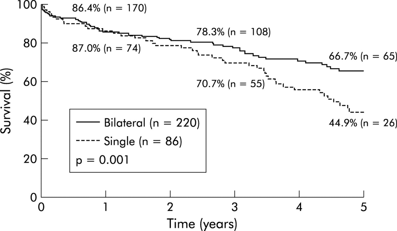
Survival estimates of 306 patients treated at Washington University with lung transplantation for a diagnosis of emphysema. Patients are stratified according to the type of transplantation performed (single v bilateral).
Table 1 shows the survival data for all lung transplants performed by our group. Patients with COPD and α1-antitrypsin deficiency emphysema make up over half the total recipients. It can be seen that patients with emphysema have a better survival rate than all other groups of patients with the exception of the 5 year survival in patients with pulmonary hypertension.
LUNG VOLUME REDUCTION SURGERY (LVRS)
The advantages of LVRS for suitable candidates are numerous, including the relief of dyspnoea and improvement in functional capabilities without the cost and adverse side effects of organ transplantation. There is no built in waiting time as with transplantation; as soon as a candidate can reach the pulmonary rehabilitation exercise goals they are ready for the procedure. The early and late mortality for LVRS are lower than those for transplantation. Without the concern for distribution of a scarce commodity such as donor lungs, lung volume reduction can be offered with slightly less rigid adherence to selection criteria. For example, a 72 year old patient who is otherwise an ideal candidate for LVRS would be considered for the procedure while such a patient would be unlikely to be added to a transplant waiting list.
The drawback of LVRS is that it is quite dependent on stringent anatomical and pathological characteristics in the patient’s lungs. Early work has shown that the lack of specific target areas and, to a lesser extent, the absence of apical target areas in particular will decrease the likelihood of a good result. Specific indications and contraindications for LVRS are shown in box 1.
Many groups have reported preliminary results for LVRS and these have consistently shown benefit to the recipient with acceptable mortality and varying morbidity.6,16–21 The remarkable finding is that these fairly uniform results have been obtained despite the use of a wide array of surgical strategies including bilateral and unilateral approaches, open and thoracoscopic operations, and buttressed or unbuttressed staplers. The consistent theme among reports of successful lung volume reduction programmes has been meticulous patient selection, methodical patient preparation with reduction of risk factors, and attentive postoperative care. Most groups have reported operating on patients with a mean age of 65 years and a preoperative FEV1 of 600–800 ml. The typical postoperative hospitalisation is 8–14 days with somewhat less than half the patients being detained for persistent air leaks from the stapled lung resection. Mortality of 0–7% has been reported for the initial hospitalisation. The expected benefits of the operation vary according to whether a unilateral or bilateral approach has been used, but gains of 20–35% in FEV1 have been reported for unilateral operations and of 40–80% with bilateral operations. Most authors also report substantial gains in exercise tolerance, freedom from oxygen use, freedom from steroid use, and subjective quality of life.
After the initial wave of single institution case series, there has been a trickle of prospective randomised trials comparing LVRS with best medical care.22–25 The results of these trials have been controversial in that they have failed to duplicate the physiological and functional gains reported in many case series. Furthermore, the mortality and morbidity in the prospective randomised trials exceeded that seen in most retrospective case series. Part of the discordance between the case series reports and the controlled trials stems from the more liberal selection criteria of the trials. By allowing patients with severe diffuse emphysema to be randomised between surgery and medical trial, the organisers of the trial by Geddes et al24 and the NETT trial22 have altered the generalisability of the results by including patients commonly felt to be contraindicated for the procedure.
COMBINATIONS OF LUNG TRANSPLANTATION AND LVRS
There are several permutations in which lung transplantation and LVRS can be combined to optimise treatment for patients with emphysema. These combinations have all been tried and have been reported in anecdotal clusters of patients. The combined approaches can be summarised as follows:
volume reduction as a bridge to transplantation;
simultaneous single lung transplantation and unilateral volume reduction to prevent native lung hyperexpansion;
early post-transplant unilateral volume reduction to treat acute native lung hyperexpansion; and
late unilateral volume reduction to treat chronic native lung hyperexpansion.
Todd et al26 recently reported the experience from Toronto with simultaneous unilateral volume reduction to prospectively improve overall lung function after a single lung transplant. They experienced no postoperative problems and the pulmonary function at 3 months was better than expected, based on historical controls receiving a single lung for emphysema. Yonan and colleagues27 retrospectively analysed 27 patients who received 31 single lung transplants for emphysema. They identified 12 patients who experienced early or late native lung hyperexpansion and they performed two early lung volume reduction operations to combat this problem. Their analysis included an assessment of risk factors and they concluded that lower pretransplant FEV1, higher residual volume, and relative pulmonary hypertension were all associated with a higher risk of native lung hyperexpansion. They did not perform or advocate volume reduction simultaneously with a single lung transplant for emphysema.
The use of volume reduction as a bridge to transplantation is the form of combined procedure that has been most frequently attempted. The concept was introduced by Zenati and colleagues28 in 1995 when they reported two patients who received single lung transplants 17 months and 4 months after laser ablation of emphysematous bullae. Bavaria et al29 prospectively performed volume reduction in patients felt also to be eligible for transplantation. They found 31 patients eligible for both procedures while, at the same time, they identified 20 patients who were suitable for LVRS alone and 139 who were felt to be candidates for transplantation only. Twenty four patients underwent successful LVRS while seven (including one death) were considered LVRS failures. The follow up was too short at the time of the report to know how frequently late transplants would be performed.
We have recently reported our results with LVRS in patients eligible for transplantation.30 Ninety nine of 200 patients who underwent bilateral LVRS were felt to have been eligible for transplantation. With a median follow up of 5.1 years, 32 of the 99 had been listed for transplantation and 15 had undergone the operation. The Kaplan-Meier curve depicting freedom from listing and freedom from transplantation is shown in fig 2. The only preoperative or operative factor that was predictive for the subsequent need for transplantation was a lower lobe rather than an upper lobe LVRS procedure.

Kaplan-Meier curve showing freedom from transplantation and freedom from listing for transplantation of 99 patients potentially eligible for transplantation after bilateral lung volume reduction surgery at Washington University.
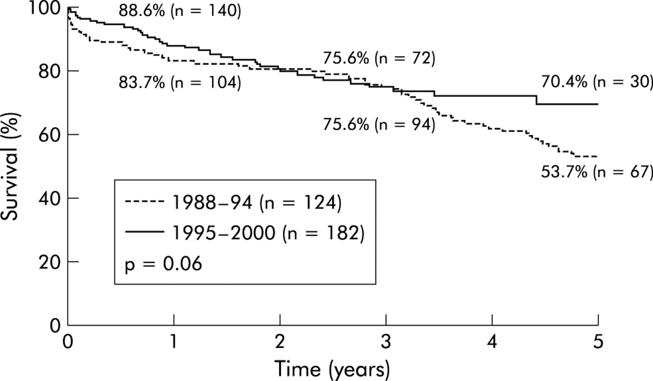
Many of our patients have had LVRS as a functional bridge to transplantation, but it has occurred not so much as part of an a priori plan to bridge them as it was additional treatment for crippling dyspnoea that was not improved sufficiently by LVRS. The concept is attractive on the surface: patients get volume reduction initially and relieve part of the demand for available lungs to transplant. The benefit for the patient who undergoes an initially successful lung volume reduction is the possibility that transplantation might be avoided altogether by an excellent response to volume reduction. A second possibility is that transplantation is delayed by several years and the patient is transplanted with a later cohort with the possibility of improved techniques, better immunosuppression, and overall better survival. Figure 3 demonstrates this effect in our own emphysema transplant recipients. Finally, since the fall off in the survival curve is steeper for lung transplant recipients than it is for those who undergo LVRS, anything that can safely delay entry onto the steeper survival curve is worth pursuing.

Survival estimates of 306 patients treated at Washington University with lung transplantation for a diagnosis of emphysema. Patients are stratified according to dates of transplantation: early experience versus recent experience.
The logic of the potential benefits of LVRS as a bridge to transplantation falls apart when faced with some aspects of reality. Firstly, the anatomical and physiological criteria for LVRS are much more restrictive than those for transplantation, so it is unlikely that a large fraction of transplant candidates could be safely and successfully treated with volume reduction. Also, the dilemma remains as to how to treat a patient near the upper age limit for transplantation. It is quite possible that a patient who is acceptable for both procedures at the age of 62 might receive volume reduction as a bridge, not to transplantation but to ineligibility for future lung transplantation several years later. Our own results have confirmed this suspicion; of the 15 patients transplanted after bilateral LVRS, only one was older than 60 at the time of evaluation for LVRS. The next oldest was 58 and the mean age for the group was 54 years.
The use of LVRS for late native lung hyperexpansion after single lung transplantation can be described as rare and anecdotal. Kroshus and colleagues reported three patients who were treated with unilateral LVRS for native lung hyperinflation and post-transplant dyspnoea that was not attributable to infection or rejection. The patients represented a small fraction of the 66 single lung transplantations performed at that centre for emphysema. The volume reduction operations were performed 12, 17 , and 42 months after the initial lung transplantation and all patients experienced a substantial relief in dyspnoea with an improvement in exercise tolerance and in the appearance of the chest radiograph.31 A similar report by Le Pimpec-Barthes et al32 describes the successful treatment of symptomatic native lung hyperexpansion by volume reduction of the native side in the form of a right upper lobectomy.
CONCLUSIONS
While bullectomy, LVRS, and lung transplantation are similar in that they each represent a surgical procedure for treating pulmonary emphysema, they are unique in their ideal selection criteria and in their expected outcomes. We favour a meticulous selection process in which all options are considered and the best option selected for a given patient. Patients referred with a functionless space occupying bulla that compresses relatively normal adjacent lung will be offered thoracoscopic or open bullectomy. The ideal indications for LVRS are hyperinflation, heterogeneous distribution of disease, FEV1 >20%, and a normal Pco2. Patients with diffuse disease, lower FEV1, hypercapnia, and associated pulmonary hypertension are directed towards transplantation. LVRS has not been a satisfactory option for patients with α1-antitrypsin deficiency and we prefer transplantation in these cases. With strict adherence to these criteria, we find that very few patients with emphysema are serious candidates for any surgical procedure. Combinations of LVRS and lung transplantation, either simultaneously or sequentially, are possible but rarely necessary.