Article Text

Statistics from Altmetric.com
- Dyspnoea
- imaging
- diagnostic
- defects
- congenital
- airway obstruction
- cough/mechanisms/pharmacology
- imaging/CT MRI
- perception of asthma/breathlessness
- rare lung diseases
A 75-year-old Caucasian man presented with a 3-month history of progressive dyspnoea, unrelated to position, with an exercise tolerance of 100 m. He had experienced thrombolysis following an inferior myocardial infarction 8 years previously.
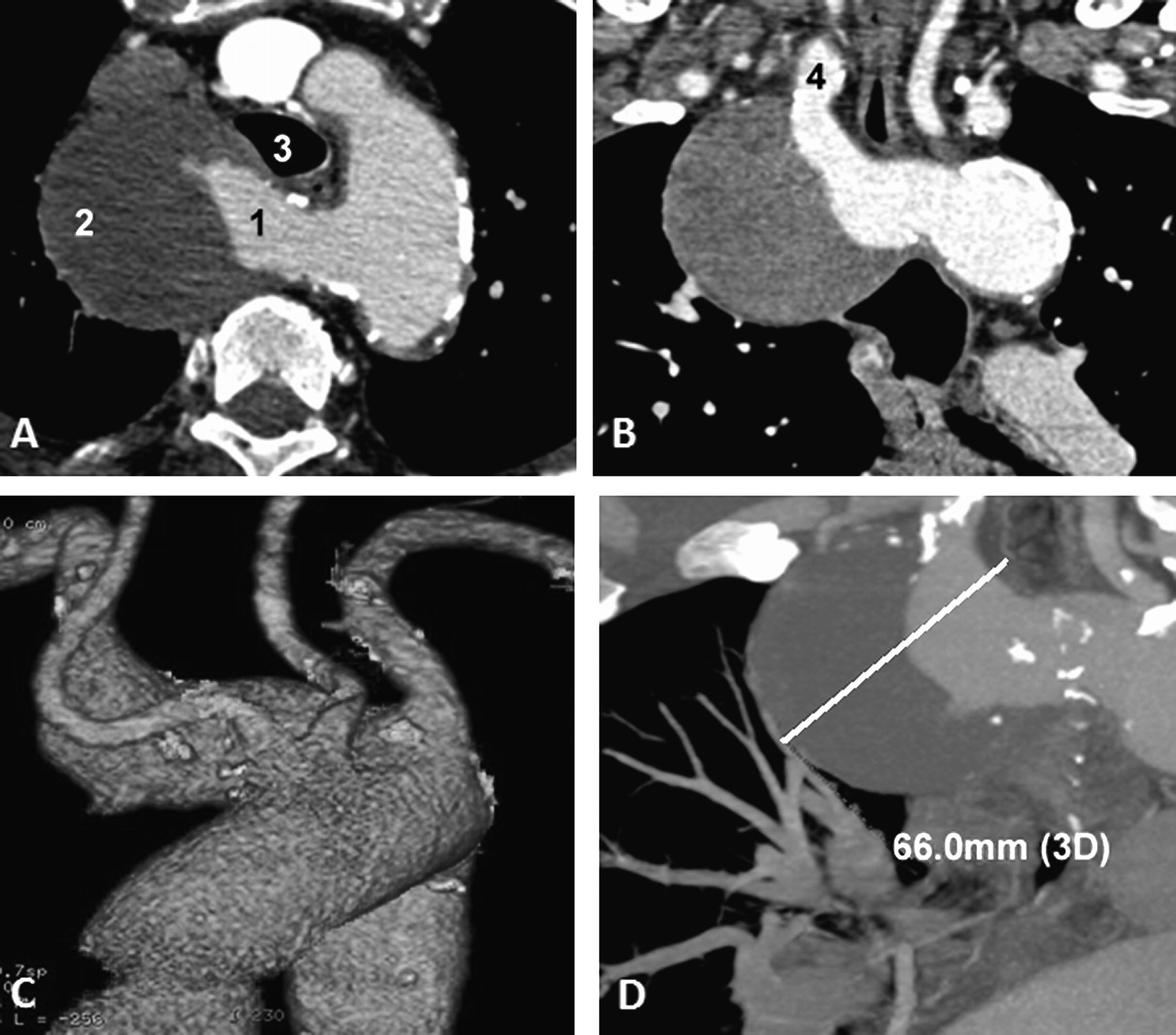
Initial diagnosis was cardiac dyspnoea; transthoracic echocardiogram showed moderate left ventricular dysfunction without significant progression from previous studies. Spirometry demonstrated fixed plateaued flow during forced inspiration and forced expiration. Cardiac MRI demonstrated an ejection fraction of 48% but no reversible ischaemia. Incidental note was made of an aneurysmal aberrant right subclavian artery compressing the trachea, further characterised by CT angiography (figure 1).

CT with intravenous contrast demonstrating an aneurysm of an aberrant right subclavian artery compressing the trachea. (A) Axial view showing the origin of the aberrant right subclavian artery; blood flow within the aneurysm (1) passes through significant intramural thrombus (2). There is partial compression of the trachea (3). (B) Oblique coronal plane showing the distal outflow beyond the aneurysm (4). (C) 3D reconstruction of the aneurysmal aberrant right subclavian artery. (D) Coronal reconstruction demonstrating the 6.6 cm maximal diameter of the aneurysmal portion.
Arteria lusoria is an anomaly occurring in 0.5–2% of individuals, but is typically asymptomatic.1 Aneurysmal dilatation may result in tracheal compression in the absence of dysphagia.
Learning points
Routine cardiac imaging techniques (transthoracic echocardiogram, coronary angiography) were insufficient to establish the correct diagnosis.
Cross-sectional imaging has a significant role to play in evaluation of patients with exertional dyspnoea whose symptoms are not explained by echocardiography.
Aneurysm of an arteria lusoria is a rare cause of obstructive dyspnoea; however, diagnosis is important to avoid the 19–53% risk of rupture.2 3
Footnotes
Competing interests The authors do not have any competing interests and/or bias with regard to this publication. All authors contributed equally to this work which has not been the subject of any preceding presentation or publication.
Patient consent Obtained.
Provenance and peer review Not commissioned; externally peer reviewed.
Linked Articles
- Airwaves