Article Text

Abstract
The relationship between allergic rhinitis and asthma is now established, and most of the clinical, epidemiological and biological data recommend integrated management. Epithelial cells represent the first barrier of the upper and lower respiratory tracts and thus are logical targets for a comprehensive integrated therapeutic approach. This review discusses rhinosinusitis as a co-morbid condition, a precipitating or triggering condition, and an epiphenomenon as an integrated part of the disease. A better understanding and a more pragmatic method of diagnosis and management is needed using cost-effective long-term strategies.
Statistics from Altmetric.com
Rhinitis and asthma have been evaluated and treated as separate disorders, but recent advances in the understanding and knowledge of the underlying processes have moved current opinion towards the concept of unifying the management of these disorders. The “united airway disease hypothesis” proposes that upper and lower airway diseases are both manifestations of a single inflammatory process.1 The upper and lower airways interface more than the air and the blood but regulate most of the human body interactions within its environment. Living an entire life with a clean and silent two-step air filter which is not replaceable suggests abilities of plasticity, regulation, teaching, renewal and local to systemic control functions. Adaptive and innate immune systems play a major role in these functions, which are centrally orchestrated within the epithelial barrier.2
Epidemiological and clinical aspects
The prevalence of asthma and rhinitis is increasing worldwide. Environmental factors are mostly thought to be responsible. Both diseases frequently coexist in the same patients, with asthma present in 20–50% of patients with allergic rhinitis and rhinitis present in up to 80% of patients with asthma.3 4 5 These data are highly dependent on the way the conditions are defined.1 Whether allergic rhinitis precedes asthma, triggers asthma or precipitates asthma are intuitive aspects that require supportive data. The atopic status plays a potentially prominent role in this relationship, but this is not a prerequisite. In a large European cohort, rhinitis was found to be an independent risk factor for asthma, not only in atopic subjects (n = 1461, odds ratio (OR) 8.1 (95% confidence interval (CI) 5.4 to 12.1)) but also in non-atopic subjects (n = 5198, OR 11.6 (95% CI 6.2 to 21.9)).6 Challenging the nose with allergens will induce the influx of inflammatory cells in the lower airways and vice versa.7 8 Treating the nose will probably, in different ways, affect lower airway inflammation.9
Definitions and clinical aspects of asthma
Asthma is defined as a chronic inflammatory disorder of the airways in which many cells and cellular elements play a role—in particular, mast cells, eosinophils, T cells, macrophages, neutrophils and epithelial cells. In susceptible individuals this inflammation causes recurrent episodes of wheezing, breathlessness, chest tightness and coughing, especially at night or early morning. These episodes are usually associated with widespread but variable airflow obstruction that is often reversible, either spontaneously or with treatment. The inflammation also causes an associated increase in existing bronchial hyper-responsiveness to a variety of stimuli.10
The fact that the definition of such a prevalent disease is complex and refers to both physiopathological key concepts as well as clinical features is important. Given the complexity of epidemiological studies to establish a causal link between two diseases such as rhinitis and asthma, it is difficult to determine whether any patient in such a study would comply with the whole definition.
In clinical practice a widespread clinical spectrum is commonly seen, as well as many co-morbidities, confounding and/or precipitating conditions including atopy, rhinosinusitis, gastro-oesophageal reflux, aspirin intolerance, exposure to cigarette smoke and occupation-related exposures.11 In this review, rhinosinusitis will be discussed as a co-morbid condition, a precipitating or triggering condition, and as an epiphenomenon as an integrated part of the disease.
Current guidelines attempt to base the management of asthma on achieving control, when previous guidelines were built on a complex step-by-step management according to clinical severity. This management is closer to the real world and will perhaps provide room for improvement in the overall control of the disease worldwide.
Non-specific bronchial hyper-responsiveness is a common finding in patients with rhinitis, even though asthma symptoms may initially be lacking. Asthma control refers to brief asthma symptoms and the absence of exacerbations. Exacerbations of asthma are events requiring unscheduled visits to healthcare providers and an important change in pharmacological management—usually a short course of oral corticosteroids. The permanent absence of control, recurrent exacerbations and high medication requirements despite optimal management define “difficult asthma”. This should be investigated using step-by-step investigations to secure the diagnosis of asthma, assess compliance issues and co-morbidities and, in some cases, will lead to the diagnosis of severe asthma.11 In these situations the role of rhinosinusitis should be carefully reassessed using clinical and imaging investigations. Rhinosinusitis is frequent and extensive in severe asthma.12 It has been shown that ethmoidal involvement is a specific feature of severe asthma, and a relationship has been found between extensive sinusitis and airway inflammation and indirect indices of distal airway changes.13
Definition and clinical aspects of rhinosinusitis
Rhinitis is defined as inflammation of the nasal mucosa characterised by nasal discharge, blockage, sneezing and itching, with two or more symptoms occurring for more than 1 h on most days. It can be further classified as intermittent (symptoms occurring on <4 days out of 7 or for <4 weeks per year) or persistent (symptoms occurring on at least 4 days out of 7 or for >4 weeks per year).14 15 The impact of chronic rhinitis on sleep, daily activities, work or school is a major determinant of impairment of quality of life in patients with asthma.3 As mentioned above, there is a significant relationship between the severity of sinus involvement and asthma severity.12 This could be related to a high level of systemic inflammation in patients with severe asthma. The perception of nasal symptoms is highly variable. This fact has been illustrated in patients with chronic obstructive pulmonary disease in whom a discrepancy has been found between nasal inflammation and symptoms.16 From a clinical point of view, it is therefore unsatisfactory to rely completely on the report of symptoms by patients as the only way of assessing rhinosinusitis.17
It is worth establishing a comprehensive biological pathway that will unify the upper and lower airways to support the epidemiological and clinical interpolations summarised above. This will lead to the development of new ways to assess the whole airway tree using non-invasive biomarkers.
Effects of anti-inflammatory treatment
Intranasal corticosteroids and antihistamines (intranasal or oral) have been shown to have anti-inflammatory effects on different aspects of inflammation in allergic rhinitis.18 Other potential treatments are intranasal cromones which stabilise mast cells by inhibiting degranulation and release of pro-mediators and newly formed mediators; intranasal anticholinergic therapy which provides relief only for excessive rhinorrhoea; and leukotriene antagonists which block the cysteinyl leukotriene receptor.19 20
In patients with asthma, inhalation of corticosteroids reduces bronchial inflammation by days or weeks, whereas reductions in airway remodelling features are reported after about 1 year.21 The clinical benefits of early treatment with anti-inflammatory treatment for asthma have been shown.22 It has been reported that the treatment of the nose may affect symptoms of asthma and bronchial hyper-responsiveness in mild allergic asthma. In some studies the use of nasal steroids was reported to be of equal value to low doses of bronchial steroids. However, asthma and rhinitis have been treated as separate diseases, but most of the current national and international recommendations (GINA, ARIA, ANAES) suggest a combined approach or at least the treatment of rhinitis as a co-morbid condition in asthma. A managed care study that included children with asthma and co-morbid rhinitis, sinusitis or otitis media showed that treatment directed at the upper airways was associated with a 30% reduction in asthma-related visits to the emergency department.23 24 Observational data in adults suggest that prescribing a treatment to control rhinosinusitis symptoms may lead to a better control of asthma and a subsequent reduction in exacerbations.25 The consequences are not only theoretical, but are important for diagnostic and therapeutic purposes.26 In patients with severe asthma with extensive pan-sinusitis, clinical reports have shown that surgical ethmoidectomy is of benefit but a placebo controlled study is difficult to undertake in this situation.
Pathological aspects
Differences and similarities between nasal and bronchial mucosa
While the nasal and bronchial mucosa have some evident similarities such as a pseudostratified epithelium and the presence of both ciliated and columnar cells resting on a basement membrane, differences are mainly seen at the submucosal level (fig 1). The large highly developed vasculature of the nose contrasts with the smooth muscle bundles that surround the bronchial airways.1 Whether inflammation-induced differential symptoms represent the expression of these anatomical discrepancies needs to be established.

(a) Nasal and (b) bronchial biopsies obtained from the same patient with mild asthma showing CD8 T lymphocyte immunoreactivity of nasal and bronchial biopsies, epithelial columnar cells, epithelial shedding and basement membrane (personal unpublished data). Original magnification ×400.
Tight junctions, peptidases and a large antioxidant apparatus are key features of the anatomical barrier of the nasal epithelium. Epithelial and endothelial permeability to small molecules (<1000 Da) in the nose is likely to produce plasma concentrations of medications close to direct intravenous administration. The mucosal associated lymphoid tissue is more developed in the nose than in the bronchi.
In asthma, epithelial fragility is a common finding while basement membrane thickness is related to the severity of the condition. In a study comparing the nose and bronchi of the same patients, eosinophilia, epithelial shedding and basement membrane thickness were similar in controls and steroid-dependent patients with asthma, while these features were more pronounced in untreated patients with mild asthma at the bronchial level (figs 2 and 3). Enhanced abilities of regeneration and protection present in the nose support the hypothesis that the nasal mucosa should be more resistant to inflammation and able to withstand environmental injuries on a near-constant basis without scarring.27 28

Epithelial thickness of nasal and bronchial biopsies obtained from controls (n = 6) and patients with mild asthma (n = 16). Epithelial thickness is decreased (epithelial shedding increased) in the bronchi compared with the nose in the patients with asthma (personal unpublished data); p<0.001 (Wilcoxon test).
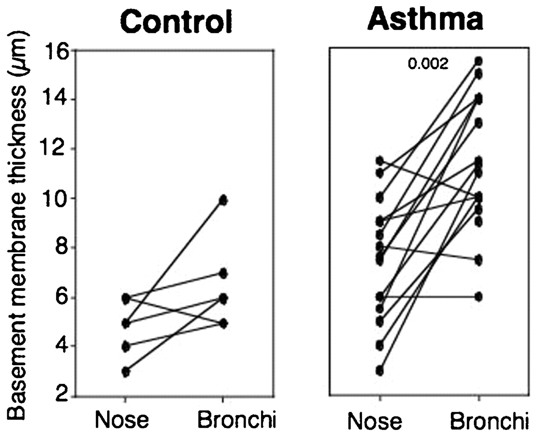
Basement membrane thickness of nasal and bronchial biopsies obtained from controls (n = 6) and patients with mild asthma (n = 16). Epithelial thickness is increased in the bronchi compared with the nose in the patients with asthma (personal unpublished data); p = 0.002 (Wilcoxon test).
Inflammatory changes seen in the nasal mucosa are closely related to those observed at the bronchial level. The same patterns of inflammatory cells such as eosinophils, mast cells, T cells and macrophages are found.29 30 Similar chemokines and cytokines are expressed at both sites, including eotaxin, interleukin 5 (IL5), RANTES, IL4, IL13 and granulocyte-macrophage colony stimulating factor (GM-CSF).7 8 Most of these molecules trigger a mainly local eosinophilic inflammatory process (fig 4).

Eosinophil number assessed as EG2 positive cells in nasal and bronchial biopsies of patients with mild asthma. The number of eosinophils in the bronchi is decreased compared with the nose in the patients with asthma (n = 16) (p = 0.01, Wilcoxon test) whereas no eosinophils were found in control subjects (personal unpublished data).
These observations support the unified airway hypothesis as the structural and inflammatory changes—both static and also dynamic after challenges—are more or less similar.
Epithelial cells are crucially situated at the external–internal milieu interface and will perform most of the actions cited above—plasticity, regulation, regeneration, orchestration of the inflammatory process and structural changes—by the production of cytokines, chemokines and other molecules balancing the inflammatory process.
Epithelial cells
The airway tree is complex and cell populations vary systematically both by airway generation and by species. In human upper airways the epithelium is pseudostratified, prismatic and ciliated, and separated from the underlying chorion by a basal membrane as in the proximal level of the lower airways. Indeed, in the large airways of the lower respiratory tract, the major cell types are ciliated, undifferentiated columnar, secretory and basal cells and also a number of less common cell types such as cartilage cells, mucus glands and neuroendocrine cells. In the small airways the cell types are similar, with relatively more ciliated cells and the secretory cells shift to the Clara cell type. At the distal level the airway epithelium merges with the alveolar epithelium, with type I and type II cells.31 The airway epithelium acts as a barrier protecting the lung from inhaled substances. Indeed, the epithelium is the point of first contact for breathable particles, respiratory viruses and airborne allergens and, as such, constitutes the interface between the external environment and the internal milieu of the lung. Under normal circumstances the epithelium forms a highly regulated and almost impermeable barrier through the formation of tight junctions.32 33 It also serves to regulate airway surface liquid volume and composition, mucus secretion and cilia beat to maintain a sterile lung through effective mucociliary clearance. In addition to its classic barrier function in response to harmful substances, the bronchial epithelium modulates immune and inflammatory responses by releasing pro- or anti-inflammatory mediators.
Mediator functions of epithelial cells
The epithelium can generate a range of products but, specifically, the synthesis and release of mediators involved in the attraction and activation of cells to the site of inflammation represents an important process in allergic mucosal inflammation and in asthma.
In nasal biopsy samples from patients with seasonal allergic rhinitis there is an increased expression of intercellular adhesion molecule 1.34 In the same way, enhanced expression of IL6, tumour necrosis factor α (TNFα), IL-8, GM-CSF and RANTES (CCL5) has been demonstrated within the airway epithelium in nasal biopsies taken from individuals with perennial allergic rhinitis.35 Eotaxin (CCL11), a chemokine also released by epithelial cells, is upregulated in the nasal tissue of patients with acute allergic rhinitis,36 and nasal challenge with eotaxin has been found to induce nasal eosinophilic inflammation. GM-CSF and stem cell factor (SCF), both generated by epithelial cells, promote survival of inflammatory cells in situ, particularly eosinophils and mast cells. Cultured nasal epithelial cells have been shown to generate SCF in vitro, and elevated levels of SCF have been found in nasal lavage fluid in patients with seasonal allergic rhinitis37 and correlated with nasal lavage mast cell chemotactic activity.
In the same way, the airway epithelium in patients with asthma is a major source of cytokines and chemokines that are strongly implicated in maintaining asthmatic inflammation, including IL8, TNFα, GM-CSF, IL5, RANTES (CCL5), eotaxin (CCL11), macrophage chemotactic peptides or thymic stromal lymphopoietin (TSLP). For example, TSLP is generated in large amounts by airway epithelial cells in response to selective toll-like receptor stimulation and shows the ability of the epithelium to provide a microenvironment sustaining ongoing Th2-like inflammation.38
Regulation of mucus secretion
The airway epithelium is also implicated in the regulation of mucus secretion by both goblet cells of the epithelial lining and mucous cells of the submucosal glands. Hypersecretion of mucus, driven by microbial factors and innate and adaptive immunity, is an integral component of the immune response to pathogens. These hypersecretory responses are seen in diseases such as asthma and allergic rhinitis and appear to be a major contributor to the severity of the condition. Indeed, in these chronic inflammatory airway diseases there is enlargement of the submucosal glands and increased numbers of goblet cells in the airway surface epithelium and their presence in the distal airways where they do not normally occur.39 40 Animal models and some human studies have described factors that contribute to goblet cell metaplasia. It is suggested that an increase in IL4 and/or IL13 participates in the process through differentiation of ciliated epithelial cells into a goblet cell/mucus-producing epithelium characteristic of Th2-mediated inflammation.41 42 43 Activation of epidermal growth factor receptors (EGFRs) on epithelial cells during repair by transforming growth factor α (TGFα) is also involved in goblet cell metaplasia.44 Finally, through activation of dual oxidase 1 (Duox1), reactive oxygen species generated by epithelial damage stimulate TGFα cleavage from its membrane precursor to promote mucus metaplasia.45 The major constituents of mucus are the mucin glycoproteins and, of the identified mucin genes (MUCs), expression of the secreted mucin MUC5AC is increased in asthmatic epithelial cells,46 47 contributing to the viscous mucus characteristic of asthma.48 At the genetic level, four highly polymorphic genes (MUC6, MUC2, MUC5AC and MUC5B) which encode gel-forming mucins play a role.49 Moreover, IL4, IL9 and IL13 induce selective mucin expression in airway epithelial cells and also the calcium-activated chloride channel 1 (hCLC A1; Gob 5), which is itself involved in regulating the expression of gel-forming mucins.50 A recent study has shown expression of nerve growth factor (NGF) in the cytoplasm of mucous cells and in a few cells of the basal layer in the nasal mucosa,51 as has been shown in the bronchi.52 Bresciani et al51 speculate that NGF produced and released in epithelial and glandular structures of the airways may play a critical role—possibly via tachykinin release—in mucus hypersecretion, a typical sign of allergic inflammation of the airways which occurs in both asthma and rhinitis. An in vitro study showed that NGF induces MUC5AC overexpression in epithelial cell lines and, similarly, a mouse model overexpressing NGF in Clara cells showed goblet cell hyperplasia and MUC5AC overexpression.
Epithelial damage in asthma and rhinitis
The airway epithelium of patients with asthma is largely abnormal, with evidence of fragility and goblet cell hyperplasia and metaplasia. This fragility is not only limited to the lower airways, since disrupted desmosome formation has also been shown in nasal polyps from patients with asthma.53 Moreover, this epithelium is also more susceptible to oxidant injury and apoptosis than normal epithelium,54 55 and it is becoming apparent that normal repair processes in the asthmatic epithelium are also compromised. Indeed, during epithelial surface injury the normal response is to upregulate receptors, notably EGFR, to drive proliferation and repair.56 However, the expression of the EGFR is increased in the asthmatic epithelium—especially in areas where columnar cells have been shed,57 but also in areas of intact epithelium—and does not correlate with the proliferative response of repairing cells.58 Furthermore, several pro-inflammatory transcription factors such as nuclear factor-κB, activator protein-1, signal transducer and activator of transcription 1 (STAT1) and STAT6 or heat-shock proteins are unnaturally expressed in the epithelium of patients with asthma.59 60 61 In allergic rhinitis the epithelial cells become activated and an accumulation of mast cells, eosinophils and basophils occurs within the airway epithelium in addition to eosinophil accumulation within the deeper lamina propria.35 In patients with allergic rhinitis without asthma, inflammatory cells and mediators have been found in the lower airways, although there is no apparent clinical manifestation of lower airway inflammation.8 62 Likewise, in the upper airways of patients with asthma, an eosinophilic infiltration has been demonstrated even in the absence of rhinitis.30
Resolution of airway inflammation
As epithelial cells have pivotal regulatory roles in inflammation and host defence against pathogens, it is also of interest to elucidate regulatory signals that could physiologically attenuate activation of epithelial and inflammatory cells both in asthma and rhinitis.
Lipoxins are lipid mediators locally produced via cell–cell interactions between leukocytes and resident cells during inflammation in human tissues to prevent an over-exuberant inflammatory response and limit damage to the host.63 In vitro and in vivo studies have shown that lipoxins display diverse potent anti-inflammatory actions including regulation of human bronchial epithelial cell function after injury to limit pro-inflammatory responses and promote a return to homeostasis.64
Lipoxin A4 (LXA4) exerts its biological actions through its specific high affinity G protein-coupled receptor, formyl-peptide receptor like-1 (FPRL-1) or ALX.65 This receptor is widely expressed in the epithelium in human proximal endobronchial biopsies.66 Lipoxins are “stop signals” for airway inflammation, acting directly on epithelial cells via interaction with its cognate receptor FPRL-1. We can postulate that upregulation of the lipoxin signalling circuits would facilitate restitution of airway epithelial homeostasis and subsequent potential resolution of inflammation, not only in asthma but also in rhinitis, and this represents a potential new therapeutic approach.
Conclusion
The relationship between allergic rhinitis and asthma is now established. Most of the clinical, epidemiological and biological data recommend integrated management. Epithelial cells represent the first barrier of the upper and lower respiratory tracts and thus are logical targets for a comprehensive integrated therapeutic approach. These diseases impact on the patient’s quality of life and are a financial burden for our healthcare systems. A better understanding and a more pragmatic method of diagnosis and management is needed using cost-effective long-term strategies.
REFERENCES
Footnotes
See Editorial, p 923
Competing interests None.
Provenance and Peer review Commissioned; not externally peer reviewed.
Linked Articles
- Editorial
- Review article
- Airwaves