Article Text

Download PDFPDF

A patchy consolidation at the apical segment of the left lower lobe associated with productive cough
Free
  1. Y-L Chang1,
  2. C-T Wu1,
  3. Y-C Lee2
  1. 1Department of Pathology, National Taiwan University Hospital and National Taiwan University College of Medicine, Taipei, Taiwan
  2. 2Departments of Surgery and Traumatology, National Taiwan University Hospital and National Taiwan University College of Medicine, Taipei, Taiwan
  1. Dr Y-C Lee, National Taiwan University Hospital and National Taiwan University College of Medicine, Roosevelt Road, Taipei, Taiwan; ntuhycl{at}gmail.com

Statistics from Altmetric.com

Request Permissions

If you wish to reuse any or all of this article please use the link below which will take you to the Copyright Clearance Center’s RightsLink service. You will be able to get a quick price and instant permission to reuse the content in many different ways.

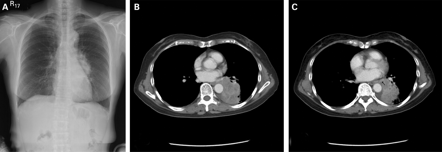
CLINICAL PRESENTATION

A 65-year-old female non-smoker had a 3-month history of productive cough and intermittent haemoptysis for 1 month. Plain chest radiography showed a mass-like shadow at the medial side of the left lower lung field (fig 1A). A CT scan showed a left lower lobe mass with heterogeneous content neighbouring the left pulmonary artery and descending thoracic aorta, with encasement of the superior and basal posterior segments of the bronchi (fig 1B). A CT scan with contrast medium revealed a highly suspicious feeding artery arising from the descending aorta (fig 1C). Bronchoscopy demonstrated narrowing of the orifice of left B6 and B10 but sputum and washing/brushing cytology were all negative. The CT-guided biopsy specimen had few atypical cells, but a repeat ultrasound-guided biopsy specimen showed chronic inflammation.

Figure 1 (A) Chest radiograph showing left lower lobe consolidation. (B) CT scan revealing a left lower lobe mass neighbouring the left pulmonary artery and descending thoracic aorta with encasement of the bronchi. (C) CT scan with contrast medium showing a highly suspicious feeding artery arising from the descending aorta (arrow).

QUESTION

What is your diagnosis?

See page 648

This case was submitted by:

Footnotes

  • Competing interests: None.

  • Patient consent: Informed consent was obtained for publication of the person's details and the figures in this report.

Linked Articles

  • Airwaves
    Wisia Wedzicha
  • Pulmonary puzzle
    BMJ Publishing Group Ltd and British Thoracic Society