Article Text

Statistics from Altmetric.com
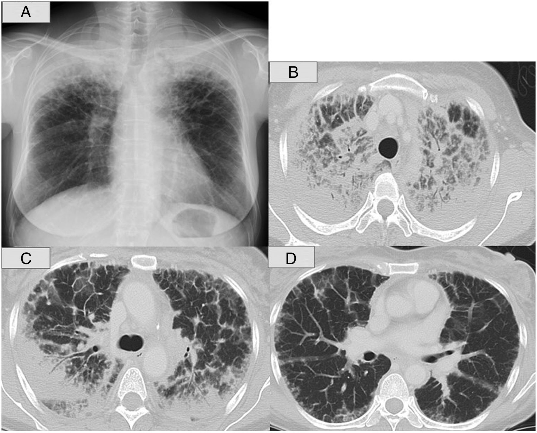
A 54-year-old female presented with chronic dry cough and dyspnoea over 3 months and was referred to our outpatient clinic. She had no history of smoking, allergy or respiratory disorders. Chest X-ray demonstrated bilateral upper lung predominant consolidation (figure 1A). Chest CT revealed extensive upper lobe predominant subpleural consolidation with air bronchograms as well as extensive ground glass opacities (GGOs) with intralobular septal thickening (figure 1B–D). Mediastinal lymphadenopathy in stations 4R and 10R with no calcification was observed. There were no crackles on auscultation with oxygen saturation of 95% on room air despite a wide range of abnormal shadows on chest X-ray and CT. Based on the available clinical and radiographic findings, the differential diagnosis encompassed pulmonary sarcoidosis, malignant lymphoma, bronchoalveolar carcinoma, lymphangitis carcinomatosa and amyloidosis.

Chest X-rays and CT scans. Chest radiograph showing bilateral upper lung predominant consolidation (A). Chest CT demonstrated extensive upper lobe predominant subpleural consolidation with air bronchograms and extensive ground glass opacities with intralobular septal thickening (B–D).
Given the diagnostic uncertainty, bronchoscopy including transbronchial biopsy was conducted and eventually facilitated a definite diagnosis. Bronchoalveolar lavage fluid (BALF) analysis of the right middle lobe demonstrated a normal proportion of differential cell counts (total cell count, 16.2×10⁴/mL in BALF; macrophages, 84.0%; lymphocytes, 15.4%; neutrophils, 0%; eosinophils, 0.6%; CD4/CD8, 2.2) with no malignant cells. Transbronchial lung biopsies from the right upper and middle lobe showed Congo red-positive amorphous eosinophilic deposits with typical green birefringence around the alveolar and blood vessel wall, suggesting pulmonary amyloidosis (figure 2A–C).

Histological findings obtained from transbronchial biopsy. Histological findings obtained from transbronchial biopsy showed amorphous eosinophilic deposits (open triangles) around the alveolar and blood vessel wall. The amorphous substances were stained by Congo red (closed triangles). Congo red stain viewed by polarising microscopy showed amorphous masses with typical green birefringence (closed arrows). (A: H&E stain, B: Congo red stain, C: polarising microscopy finding).
Blood and urine tests to detect any underlying disorders showed elevated sialylated carbohydrate antigen KL-6 of 613 U/mL (normal range <500 U/mL) and sIL2R of 631 U/mL (normal range <466 U/mL), decreased immunoglobulin (IgG 660 mg/dL; IgA 12 mg/dL; IgM 12 mg/dL) with high levels of IgG λ-type M-protein and positive Bence-Jones protein. Tumour markers (carcinoembryonic antigen, progastrin releasing peptide and cytokeratin 19 fragment) and angiotensin converting enzyme (ACE) levels were normal. Bone marrow aspiration was also performed and immunohistochemical staining revealed CD138-positive plasma cells. On the basis of the above-mentioned findings, the patient was diagnosed with diffuse alveolar septal amyloidosis associated with multiple myeloma (IgG λ-type). Autologous stem cell transplantation after a combination treatment of cyclophosphamide, bortezomib and dexamethasone was performed. At present, the patient's multiple myeloma is in complete remission, but her radiographic findings persist despite the haematological response.
Previous review papers have reported that the pulmonary manifestations of amyloidosis are mainly classified into three distinct patterns: tracheobronchial, nodular and diffuse alveolar septal patterns.1 The proportions of those radiographic patterns have been identified to be 53%, 44% and 3%, respectively,1 indicating that the present case showing alveolar septal pattern is extremely infrequent. This case is clinically significant because the majority of cases are associated with underlying systemic disorders such as multiple myeloma.2 The prognosis of pulmonary amyloidosis accompanied by multiple myeloma is poor when compared with primary pulmonary amyloidosis. Additionally, radiographic improvement of the lung after treatment is rare.3 Therefore, treatment for the primary disorder should be started immediately.
In conclusion, we presented a rare case of pulmonary amyloidosis, where radiographic observations clearly corresponded to pathological findings. The radiographic findings of consolidation and GGOs with alveolar septal thickening may warrant consideration of a diagnosis of pulmonary amyloidosis.
Acknowledgments
We thank Dr Osamu Suzuki, Department of Diagnostic Pathology in Fukushima Medical University, for providing us with images of histopathological specimens, including Congo red staining with typical green birefringence.
Footnotes
Contributors YS, JS, RT, HM and MM were in clinical care of the patient; YS, JS and MM wrote the manuscript.
Competing interests None.
Patient consent Obtained.
Provenance and peer review Not commissioned; externally peer reviewed.