Article Text

Statistics from Altmetric.com
A 62-year-old man diagnosed with glioblastoma multiforme and currently under chemoradiation therapy presented with massive haemoptysis. Chest computed tomographic scan disclosed a round-shaped formation in the right upper lobe with contrast agent leakage indicating active bleeding (figure 1). Pathological examination after right upper lobectomy revealed a pseudoaneurysm of the pulmonary artery caused by invasive mucormycosis (figure 2).

Contrast-enhanced computed tomography of the chest showing the pseudoaneurysm of the right upper lobe pulmonary artery. Image also demonstrates a contrast agent leakage indicating an active bleeding.

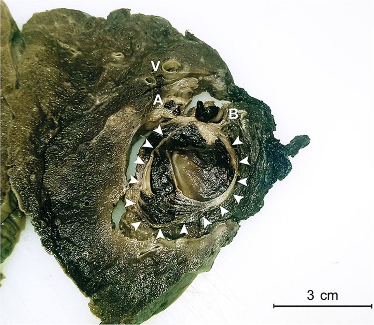
Resected right upper lobe with pseudoaneurysm (arrows) of the pulmonary artery (A). B, bronchus; V, vein.
After candidiasis and aspergillosis, mucormycosis is the third most frequent invasive mycosis in immunocompromised patients. In 25% the lungs are the main site of manifestation and in these cases a high mortality is reported.1 An interesting characteristic of Mucorales species is the angioinvasion which leads to rupture of vessels with massive haemorrhage.2
Footnotes
Competing interests None declared.
Patient consent Obtained.
Provenance and peer review Not commissioned; internally peer reviewed.