Article Text

Statistics from Altmetric.com
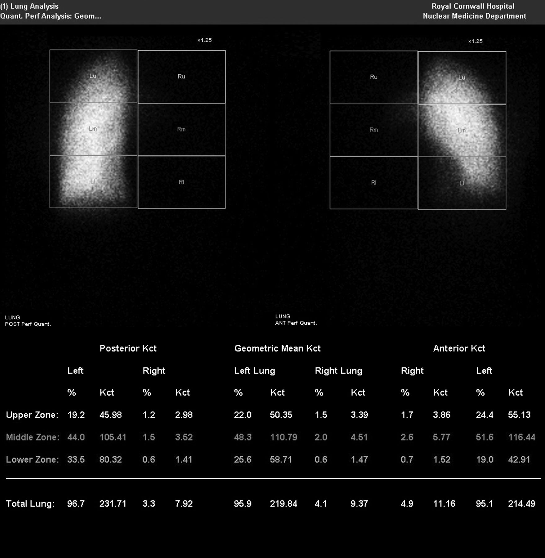
An 18-year-old female subject with a history of decortication for right sided, postpneumonic pleural thickening presented with haemoptysis. A CT pulmonary angiogram showed marked hypoplasia of the right main, lobar and segmental pulmonary arteries, along with reduced volume of the right lung. Prominent bronchial artery supply to this lung was noted (figure 1) with absence of pulmonary veins. Perfusion scintigraphy showed only 4% perfusion to the right lung and normal perfusion of the left lung (figure 2). Pulmonary artery hypoplasia has been reported to be associated with ipsilateral pulmonary venous atresia.1 It can present with symptoms mimicking pulmonary embolism.2
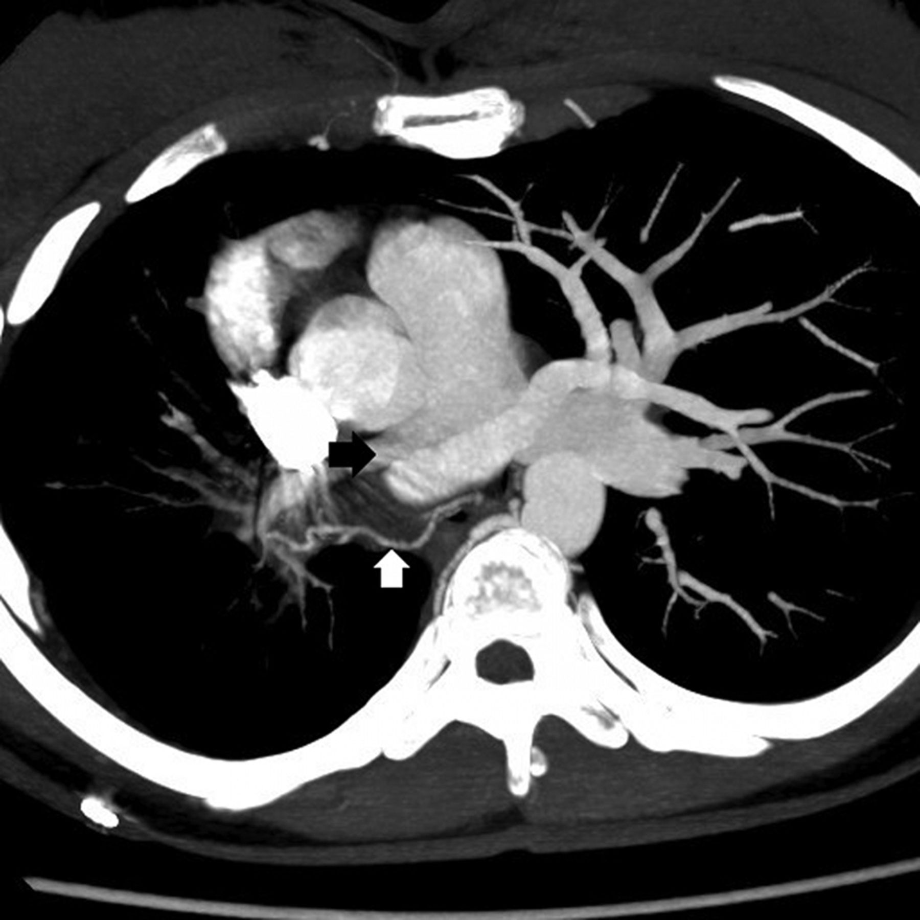
Axial maximum intensity projection image from CT pulmonary angiogram showing hypoplastic pulmonary artery (black arrow) and prominent bronchial artery (white arrow) on the right side.

Perfusion scintigraphic images demonstrating scant perfusion to the right lung.
Learning points
-
Pulmonary artery hypoplasia is a rare cause of haemoptysis. The source of bleeding is usually from hypertrophic bronchial arteries.
-
CT pulmonary angiogram combined with perfusion scintigraphy is very useful in making the diagnosis non-invasively.
Acknowledgments
Mr Ivor Dixon, Medical Physicist, Royal Cornwall Hospital, Truro, Cornwall, UK for providing the new figure.
Footnotes
-
Competing interests None.
-
Patient consent Obtained.
-
Provenance and peer review Not commissioned; internally peer reviewed.
Linked Articles
- Airwaves