Article Text

Statistics from Altmetric.com
A 60-year-old man was admitted under the cardiology team for increasing shortness of breath over the past two months. His medical history of note included stable renal transplant 8 years ago, previous parathyroidectomy due to uncontrolled tertiary hyperparathyroidism and hypertension. His echocardiogram showed a critical aortic stenosis and was thus worked up for an aortic valve replacement.
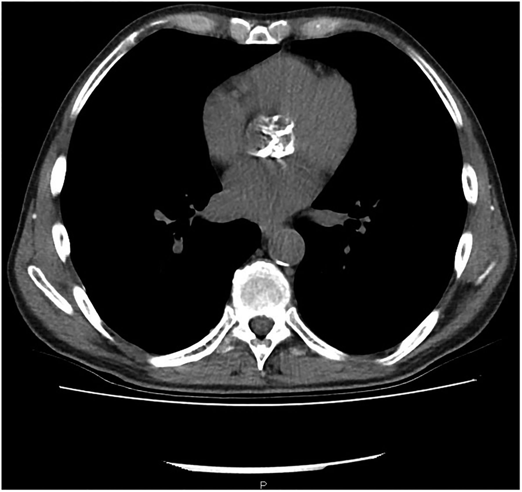
His admission X-ray (figure 1) showed bilateral apical nodular alveolar opacities. High-resolution CT chest is as shown in figure 2 and was described superficially like a ‘crazy paving’ pattern by the reporting radiologist (figure 2). There is also severe calcification of the aortic valve noted (figure 3).

Chest X-ray depicting bilateral apical nodular alveolar opacities.
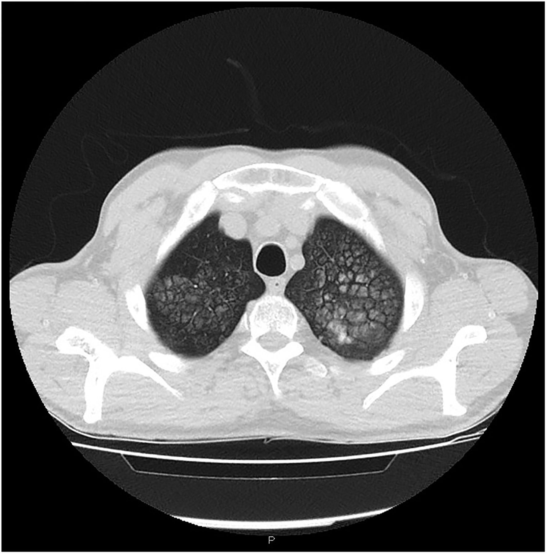
High-resolution CT chest showing calcification of both lung apices.

Same CT scan showing a heavily calcified aortic valve, most probably related to his previous hypercalcaemia.
Metastatic pulmonary calcification is seldom recognised clinically as it is usually asymptomatic. High-resolution CT scan is sensitive and specific as it is able to depict small amounts of calcification seen compared with conventional chest X-ray.1 Upper lobes are mainly involved (as in this patient) because of the high ventilation-perfusion ratio, which results in lower capillary pCO2 and a higher pH compared with the lower lobes.2 It is important to know that despite the term ‘metastatic’ being used, it is a relatively benign condition with a good long-term prognosis.
Acknowledgments
The author acknowledges the contribution of Dr Paul Grech, the radiologist responsible for reporting the findings of the high-resolution CT scan.
Footnotes
Competing interests None declared.
Patient consent Obtained.
Provenance and peer review Not commissioned; internally peer reviewed.
Linked Articles
- Airwaves