Article Text

Abstract
A 19-year-old boy with shortness of breath and chest pain after strenuous exercise presented to emergency department . On physical examination, the neck and shoulders appeared to be swollen. There was crepitus on skin palpation. Chest X-ray disclosed diffuse subcutaneous emphysema and pneumomediastinum. CT showed additional finding of air in epidural space. The patient was discharged after 2 days of hospitalisation with conservative treatment uneventfully. Pneumorrhachis is usually caused by abrupt increase in intrathoracic pressure in instance of forceful vomiting, cough or asthma attack in an otherwise healthy young adult. It is usually accompanied with pneumomediastinum. The management of epidural pneumatosis should be tailored according to its primary cause. For most patients with pneumorrhachis associated to a spontaneous pneumomediastinum without neurological symptoms, this condition is generally self-limited. For epidural free air of large volume that causes neurological deficits, surgical laminectomy may be indicated.
- Asthma
Statistics from Altmetric.com
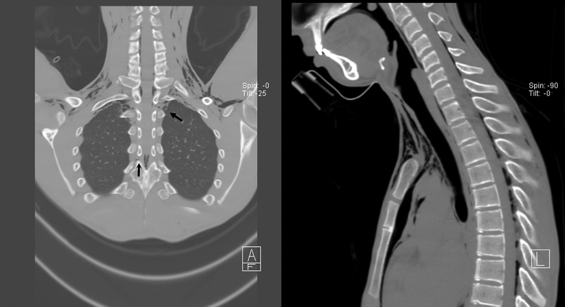
A 19-year-old boy with shortness of breath and chest pain after strenuous exercise presented to emergency department (ED). He also had a common cold for 1 week prior to ED visit. On triage, he was afebrile, and haemodynamics were stable. On physical examination, the neck and shoulders appeared to be swollen, revealing crepitus on skin palpation. Chest X-ray disclosed diffuse subcutaneous emphysema and pneumomediastinum. CT showed additional finding of air in epidural space (figure 1). The patient was discharged after 2 days of hospitalisation with conservative treatment uneventfully.

CT of the chest, showing extensive subcutaneous emphysema, pneumomediastinum and epidural pneumatosis. Free air dissected along the intervertebral foramens of upper thoracic vertebrae (arrow).
Pneumorrhachis is usually caused by abrupt increase in intrathoracic pressure in instance of forceful vomiting, cough or asthma attack in an otherwise healthy young adult. It is usually accompanied with pneumomediastinum, since the air from pulmonary alveoli has to travel across the mediastinum in order to reach the epidural space.1 It is estimated that pneumorrhachis occurs in 9.5% of patients with spontaneous pneumomediastinum, which is already a rare disease entity and is responsible for 1 of 8005 to 1 of 42 000 hospital incidents and emergency admissions.1 ,2 Other possible origins of free air include retropharyngeal space, illicit drug use or direct trauma to the vertebral column. The presence of epidural free air usually does not cause symptoms; however, neurological deficits may occur if the volume of free air is large.3
The management of epidural pneumatosis should be tailored according to its primary cause. For most patients with pneumorrhachis associated with a spontaneous pneumomediastinum without neurological symptoms, this condition is generally self-limited if cough, emesis or asthma is under control. For pneumorrhachis of other aetiologies, such as oesophageal rupture, emphysematous infection and open trauma of the spine, more aggressive and specific treatment should be undertaken. For epidural free air of large volume that causes neurological deficits, surgical laminectomy may be indicated.
Footnotes
Contributors P-YL is the main writer and primary care physician of emergency department, and contributed to patient history. H-JW was the reporting radiologist and contributed to several parts of discussion.
Competing interests None.
Patient consent Obtained.
Provenance and peer review Not commissioned; internally peer reviewed.