Article Text

Statistics from Altmetric.com
The birth of the single breath (sb) TLCO: the oxygen secretion controversy
Whether or not the lungs actively secreted oxygen, particularly under stressful conditions (severe exertion, alveolar hypoxia), was an argument which continued for more than 50 years (1870–1923), and involved some of the most distinguished respiratory physiologists of that era, such as JS Haldane, Christian Bohr and August Krogh. The denouement, as told by Krogh's daughter, Bodil Schmidt-Neilsen,1 began 11 years earlier in 1904 when a Danish medical student, Marie Jørgensen, attended a class taught by August Krogh, an instructor in physiology in Christian Bohr's department. They were attracted to each other and married in 1905, and Marie (figure 1) joined August (a Nobel Prize winner in physiology and medicine in 1920) in some aspects of his research. The subsequent publication of a paper in 1915, 100 years ago this year, by Marie Krogh in the Journal of Physiology2 was the pivotal moment in the story. The influence of this paper “The diffusion of gases through the lungs of man”2 continues to this day, long after the oxygen secretion question was settled (in Marie Krogh's favour). Nowadays, pulmonary function laboratories throughout the world use a modification of her single breath transfer factor for carbon monoxide (TLCO.sb) test, known in North America as the DLCO.sb (carbon monoxide diffusing capacity); it is an essential part of routine lung function screening, and the only non-invasive test (apart from pulse oximetry) of the gas exchanging efficiency of the lung.

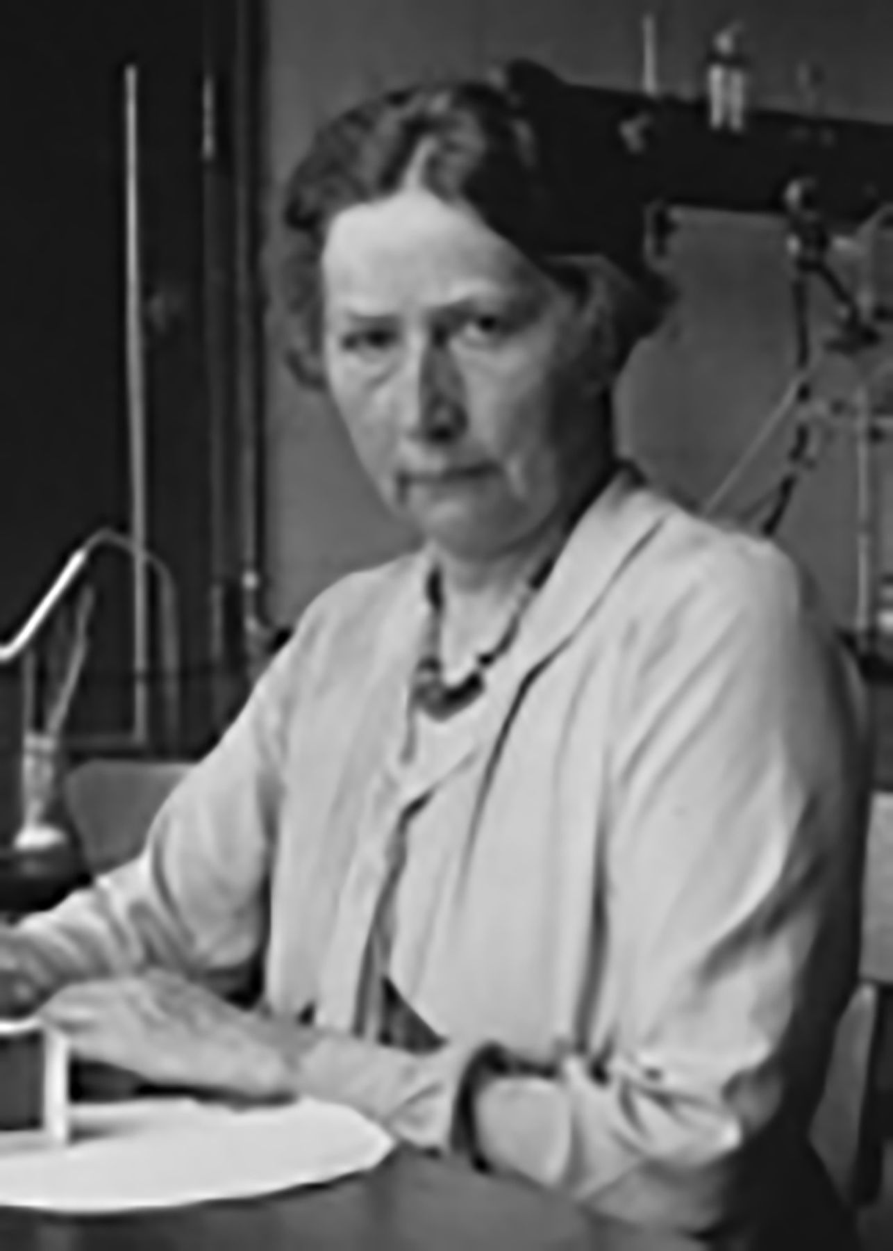
Marie Krogh (née Jørgenson), born Christmas day 1874 on the island of Fyn, Denmark, married August Krogh in Copenhagen, March 1905, died 1943 in Copenhagen, aged 69 years.
The oxygen secretion story has been told many times.1 ,3 ,4 A century ago, the idea that Claude Bernard's milieu intérieur might be stabilised by alveolar cells secreting oxygen at times of great demand or shortage would have seemed not unreasonable. There was, after all, the example in nature of O2 partial pressures ( ) in the swim bladders of fish many times higher than the environmental oxygen. Recently, however, Scheid et al5 have shown that this can occur without active secretion, due to a combination of mechanisms, (A) lactic acid production, (B) reduced haemoglobin affinity and capacity for O2, (C) a countercurrent rete mirabile, and (D) a swim bladder wall made impermeable by guanine crystals. In fact, the only evidence ever produced in favour of alveolar oxygen secretion was the finding that arterial
  was greater than alveolar
  . This occurred in the years 1890–1912 when methods of measuring
  in blood were relatively crude and inaccurate. August Krogh first solved the technical problem of measuring arterial
  accurately;6 the Kroghs found alveolar
  was always greater than arterial
  .7 But, the proponents of the secretion theory could always argue that end-capillary
  (after subtraction of the contributions from hypoxaemic blood from intrapulmonary and extrapulmonary shunts) might, on severe exercise, have been up to 1 kPa (1–7 mm Hg) above mean alveolar
  (which might have been elevated by contamination with dead space
  ). Thus, an alternative approach was needed.
When CO is inhaled, its great affinity for haemoglobin (Hb) in blood (×230 vs O2) means that its partial pressure in blood (PCO) stays negligible. The transfer factor for CO (quantity taken up per unit time, per unit PCO), or TLCO, equals the rate of uptake (ΔCO/Δt) from alveolar gas (which is easily measured) times the alveolar volume (VA) at which the measurement is made, divided by the total dry gas pressure (barometric minus water vapour pressure at 37°). The Kroghs measured the TLCO in normal subjects at rest and on exercise;8 finally, Marie Krogh repeated the measurements with a much improved method.2 Using an O2/CO diffusivity ratio of 1.23 (based on their physical properties), she calculated  from the measured TLCO, and showed that, with reasonable values for the alveolar-mean capillary
  gradient multiplied by the exercise
  (
( ), the measured oxygen consumption (
 ) of 2.6 L min−1 at the exercise levels studied, could be accounted for solely by passive diffusion. Using the principle of Occam's razor, there was no need to invoke an additional mechanism.
The 1915 single breath DLCO (TLCO)
To measure the diffusing capacity (transfer factor) for CO, only alveolar PCO and CO uptake ( ) had to be measured. How best to do it? The Kroghs tried first a steady state technique8 (like the measurement of
  ) but abandoned it because
  measurements were not reproducible during tidal breathing; a breath hold technique proved more satisfactory.2 For the same reasons, when the
  was ‘rediscovered’ in the 1950s, the single breath technique became the preferred choice over the steady state.9 The 1915
  differs from the present day
  in some technical details (see table 1), but, quantitatively, Marie Krogh's measurements have been confirmed by later investigators (table 1, note ¶).
Comparison of TLCO.sb methods since Marie Krogh's original description; shading indicates no change from the earlier date
Nomenclature and calculations in 1915
DLCO (the term “transfer factor, TLCO”, did not come in until 196314) was diffusion constant, and k, the rate of CO uptake (rate constant) per unit PCO (now referred to as  or KCO) was permeability. Marie Krogh2 states “The diffusion is determined by two factors, namely the permeability (k) and the mean capacity (∼VA)”; the relationship TLCO=KCO×VA is what we teach today.15 She also says (p. 288)2 “Two persons may have about the same diffusion constant, …. though both mean capacity and the permeability are very different”. See table 3 in a recent review.15
1923–1957: understanding the physiology of CO uptake
In 1915, Marie Krogh2 said “……. an essentially indifferent gas, like carbon monoxide, must pass through the alveolar epithelium by diffusion alone ….”. This was, at the time, a generally held assumption.4 The rate of combination of the Hb in the red cell with oxygen, and especially CO, was thought to be instantaneous. In the 1920s, with improved analytical techniques, Hartridge and Roughton in Cambridge (UK) were able to measure the rate of association of CO with solutions of Hb. They showed that the reaction velocity of CO was not instantaneous, but measureable, and that its rate of combination with Hb packed inside red cells was significantly slower than in Hb solutions, implying diffusion as well as reaction resistance to CO uptake within pulmonary capillary blood. Furthermore, the reaction resistance was proportional to red cell  . Finally, the TLCO.sb was found to be directly related to alveolar
  (note 1/TLCO is a resistance, TLCO is a conductance).4
This era of physiological discovery culminated in the formulation of the famous Roughton–Forster equation,16 which partitioned the alveolar uptake of CO into membrane (DM) and red cell (θVc) components: 1
1
where 1/DMCO is the diffusion resistance from the epithelial surface to the red cell membrane and 1/θVc is the red cell resistance to CO uptake, where θ is rate of CO uptake per mL blood (inversely proportional to  ) and Vc is the pulmonary capillary volume. Subsequently, physiological studies, involving simultaneous nitric oxide (NO) and CO uptake, have shown that 80% of the resistance to CO transfer from alveolar gas to intracapillary Hb (∼1/TLCO.sb) lies in the red cell itself.17 ,18 Thus, it is not unreasonable to describe the TLCO as a ‘window on the pulmonary microcirculation’.19
TLCO 1945–1957
There was no clinical follow-up after Marie Krogh's pioneering 1915 publication, until the TLCO (∼DLCO) reappeared in 1957, slightly modified, in a paper by Ogilvie et al9 from the University of Pennsylvania, USA. Colin Ogilvie was an English chest physician. The invention of the infrared CO meter in Germany in the early 1940s made CO analysis quicker and more practical. In addition, interest in lung diffusion had been revived in 1945 by a challenging paper from Lilienthal et al20 in which the oxygen diffusing capacity ( ) was measured. Their method was complex and involved breathing hypoxic gas mixtures (13% O2). At around this time (1949–1951), clinicians had seen patients with lung fibrosis and small lungs with a decrease in arterial O2 saturation (SaO2)>10% on exercise, from almost normal values at rest.21 Austrian et al22 measured a low
  (at rest) in similar patients. They described this as ‘alveolar–capillary block’; we now call it ‘diffusion limitation’. When resting DLCO is <60% predicted in patients with interstitial lung fibrosis, worsening of arterial hypoxaemia on exercise is extremely common.
Seymour Kety,23 a circulatory physiologist, in a review of methods for measuring pulmonary diffusion, realised that Marie Krogh's use of CO ‘brilliantly sidestepped’ the difficulties involved in measuring  . Julius Comroe in Philadelphia assembled a team, led by Robert Forster, to repeat Krogh's work with clinical application in mind. Ward Fowler, working in Comroe's department on alveolar gas distribution, made the novel suggestion of adding an insoluble ‘volume marker’ gas (helium) to the inspired mixture. This, cleverly, eliminated the first expiration (A) because the initial alveolar CO concentration (at t=0) could be calculated from the helium expired at the end of the breath hold as
  ×Hee/Hei where e and i or I refer to expired and inspired, respectively (see table 1). Thus, Krogh's first expiration was avoided, the breath hold was maintained at a reproducible level (∼TLC) and only two samples (from the inspired bag and the expired sample) had to be analysed. There had to be an assumption that the inspired CO and helium mixed instantaneously with alveolar gas; while this was unlikely to happen, the preceding fast inspired vital capacity would minimise mixing delays. This was the only modification of substance made to Krogh's method. Other modifications to Krogh 1915, made in 1957 and subsequently, are listed in table 1. The two most important are (A) standardisation of the calculation of breath hold time,11 and (B) the substitution of a helium dilution VA, measured during the single breath manoeuvre, for a separately measured residual volume to which the inspiratory vital capacity was added. This saved Pulmonary Function Laboratories from making two separate measurements. But, in the presence of airflow obstruction, it means that the ‘VA’ and the TLCO calculated from it are weighted towards the better ventilated lung regions.
TLCO·SB in 2015
Pulmonary function testing did not have a role in medicine in 1915, and its profile did not rise until the late 1940s and 1950s. Marie Krogh can hardly have foreseen that her single breath method, using CO, designed to answer a specific physiological question, would become, 50 years later, a day in, day out test in Pulmonary Function Laboratories worldwide, playing a key role in functional assessment in respiratory and systemic disease. Clinically, the TLCO.sb (and KCO) are normal in uncomplicated asthma, and abnormal in emphysema, where there is a good correlation with CT scanning indices of lung destruction.24 In restrictive lung disease, ‘small lungs’, KCO is considerably greater than predicted normal (>120%) in extrapulmonary causes, but <100% predicted with intrapulmonary pathology.15 Interpretation of a reduced TLCO.sb in disease is helped by consideration of its two separate components (KCO and VA),15 to which Marie Krogh first drew attention.2
TLNO·SB the future
In 1987–1989, two papers were published25 ,26 (although the earlier paper26 was preceded by the work27 described in the later publication25) in which gaseous NO replaced CO, and the single breath transfer factor for nitric oxide (TLNO) emerged; the theory and methodology were similar to the TLCO. These two groups, from Cambridge, UK17 and Bordeaux, France,18 have since shown that, for NO, 35–40% only of the transfer resistance lies in the red cell (80% for CO). Thus, TLNO.sb is more weighted to the alveolar and capillary surface area, that is, true diffusion, than to the red cell. So, TLNO differs from TLCO, but complements it. Furthermore, by using laboratory derived values for θCO and θNO, DMCO and Vc can be calculated from one single breath manoeuvre. The future may tell us whether the combination of TLNO and TLCO will prove more useful clinically than TLCO alone.
Acknowledgments
The authors thank Dr Harry Gribbin for the reminder of the anniversary of Marie Krogh's 1915 paper.
References
Footnotes
-
Competing interests None.
-
Provenance and peer review Not commissioned; externally peer reviewed.