Article Text

Statistics from Altmetric.com
Air embolism is the entry of air into the vascular system and is usually associated with surgeries, trauma and central lines.1
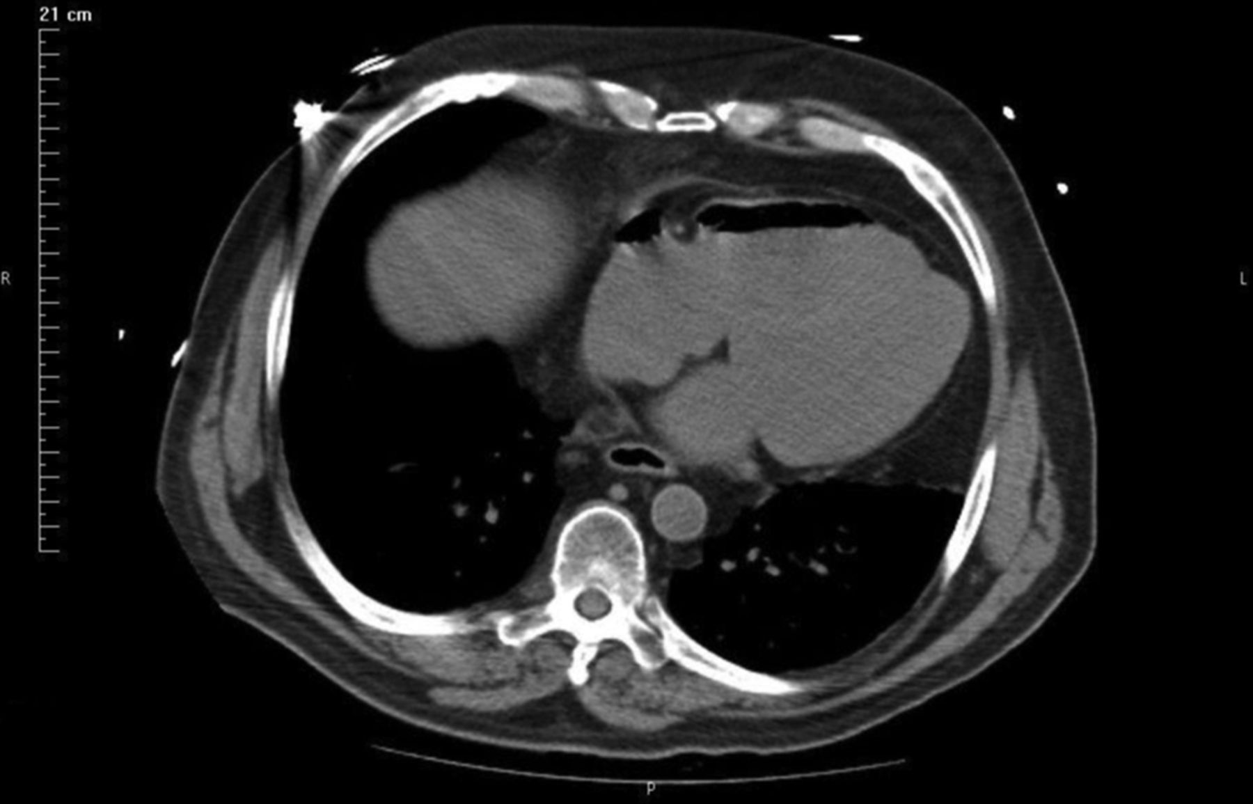
A patient presented to the clinic with new onset atrial flutter and lower extremity oedema. The patient was admitted and within 24 h, the course became complicated by respiratory failure, requiring intubation and admission to the intensive care unit. A chest CT scan with contrast was ordered for evaluation. Upon acquisition of the images, the patient was found to have at least 50 cc of air to be present in the right atrium and ventricle (figure 1), along with bilateral pulmonary embolism, the latter of which was thought to be the cause of the clinical presentation. The patient did not have any known risk factors for air embolism that is, surgery, trauma or central venous accesses. Upon close investigation, the aetiology was presumed to be iatrogenic from the use of a power injector for injecting dye for the initial CT scan. It was decided to follow this up with a repeat chest scan before taking any steps to try to remove the air. The patient, in the interim, was started on heparin for the pulmonary embolism and managed medically. A repeat chest CT scan after 12 h showed spontaneous resolution of the air (figure 2).

CT image demonstrating air present in the right atrium and ventricle.

CT image showing resolution of air in the right heart.
Air embolism can occur with the use of dye power injectors, however such large volumes are rare and may be related to critical clinical errors. This should be thought of especially in cases where no known cause of air entry into the venous system is identified.
Reference
Footnotes
-
Contributors SJ: idea conception, write-up, image acquisition. JAP: write-up and guidance/mentorship.
-
Competing interests None.
-
Provenance and peer review Not commissioned; externally peer reviewed.
Linked Articles
- Airwaves