Article Text

Abstract
Introduction Endobronchial Ultrasound (EBUS) allows minimally-invasive hilar and mediastinal lymph node sampling and has an established role in the diagnosis and staging of lung cancer. Molecular biomarker development is becoming increasingly relevant in lung cancer management, however the suitability of EBUS-derived aspirates for detailed molecular analysis is not fully defined. Gene expression profiling (GEP), a powerful micro-array technology, which assesses genome-wide changes in gene expression, can generate individual-specific molecular signatures that can provide prognostic information and predict treatment responsiveness. Here we demonstrate the feasibility of using EBUS-derived cytological aspirates from benign and tumour infiltrated lymph nodes in patients with NSCLC for GEP.
Methods Cytological aspirates from six patients with known NSCLC that had been referred for EBUS to stage the mediastinum were selected for GEP. Three patient samples were infiltrated by NSCLC and three were benign. NSCLC-infiltrated and benign lymph nodes were compared for differences in gene expression.
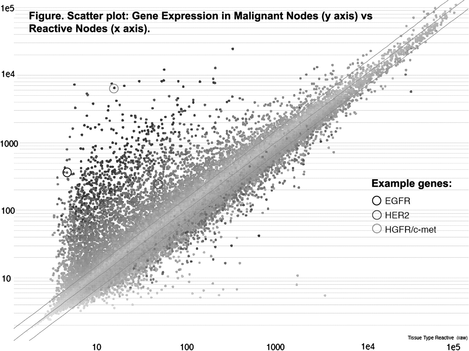
Results RNA was available at a yield (median 17.5 μg, range 0.7–62.3 μg) and integrity (RIN Median 7.1, range 5.3–8.0) suitable for amplification and GEP. Reactive and malignant nodes were differentiated by principal component analysis and hierarchical clustering with ability to identify upregulation of cancer specific genes in malignant relative to benign nodes (notably EGFR, HGFR/c-met and HER2 were among genes most upregulated).
Conclusion We demonstrate the feasibility of RNA extraction and GEP on EBUS-derived lymph node cytological aspirates and show differences in gene expression profiles between benign and tumour-infiltrated lymph node mRNA. Further studies on larger patient cohorts are necessary to identify expression profiles that can robustly differentiate benign from malignant lymph nodes in NSCLC.
Scatter plot: Gene expression in malignant nodes (y axis) vs reactive nodes (x axis).