Article Text

Statistics from Altmetric.com
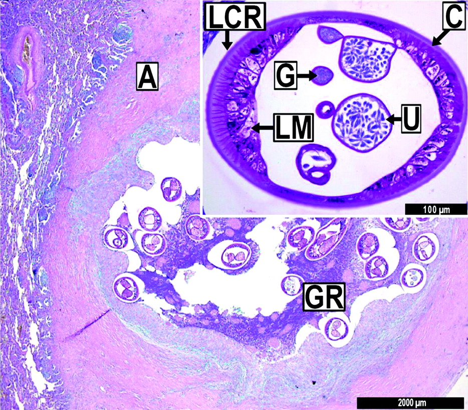
A 22-year-old Caucasian man with a history of metastatic chondroblastic osteosarcoma was wedge resected via open thoracotomy after surveillance follow-up in 2009 revealed suspicious multifocal bipulmonary lesions on a CT scan (figure 1). Five years previously, neoadjuvant and adjuvant chemotherapy according to the COSS 96 protocol1 had been given. Surveillance follow-up in 2009 showed new multifocal partly calcified pulmonary lesions in both lower lobes. Histologically, the resected lesion in the lower right lobe was vital tumour metastasis whereas the lesion in the lower left lobe showed a non-malignant nodular granulomatous reaction to Dirofilaria repens (figure 2).

CT scan in lung window showing a solid pulmonary node (SPN) surrounded by scar tissue in the left lower lobe (LLL). This lesion presumably contained the nematode Dirofilaria repens.
Periodic acid-Schiff-stained tissue sample of an embolised artery (A) containing sections of a female Dirofilaria repens (U, uterus) with its multilayered cuticle (C) and external longitudinal cuticular ridges (LCR), longitudinal muscles (LM) and gut (G). The nematode is surrounded by a granulomatous reaction (GR). Magnification ×10; inlay ×40.
Dirofilariasis is a common vector-borne zoonosis. Occasionally, nematodes are transmitted by mosquitoes to subcutaneous tissues of humans.2 In extremely rare cases such as extended periods of immunosuppression, worms migrate to the lungs and cause asymptomatic granulomatous coin lesions.2 3 Microscopically, typical pulmonary nodules display a central thrombosed artery and the parasite in various stages of degeneration, often surrounded by eosinophilia.4 To our knowledge, this is the first report of possible indigenous pulmonary dirofilariasis accompanying and mimicking lung metastasis in a patient with osteosarcoma.
Learning points
Vector-borne transmissions of Dirofilaria repens occur in humans.
Extended periods of immunosuppression can facilitate pulmonary dirofilariasis.
Pulmonary dirofilariasis is a rare differential diagnosis of tumour-like lesions.
Acknowledgments
The authors thank Dr Sven Poppert, Bernhard Nocht Institute for Tropical Medicine, Hamburg, Germany for confirming the diagnosis of Dirofilaria repens by molecular biological methods.
Footnotes
RW and TS share the senior authorship of this manuscript.
Competing interests None.
Patient consent Obtained.
Provenance and peer review Not commissioned; externally peer reviewed.