Article Text

Statistics from Altmetric.com
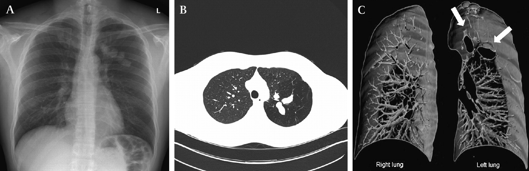
A 31-year-old, HIV-positive man presented with a history of chronic dry cough. Chest x-ray showed a rounded, branching opacity in the left upper lung (finger-in-glove sign, figure 1A). CT showed mucoid impaction, segmental hyperlucency and decreased vascularity of the left upper lobe (figure 1B). 3D reconstruction of the bronchial tree revealed an atretic apicoposterior segmental bronchus of the left upper lobe confirming the diagnosis of congenital bronchial atresia (figure 1C).

(A) Postoanterior radiograph showing a branching opacity in the left upper lung (finger-in-glove sign). (B) Axial CT image with tubular opacity and adjacent hyperlucency in the left upper lobe. (C) 3D-reconstruction of the bronchial tree. There is no division of corresponding bronchi, indicating atresia (see arrows).
Congenital bronchial atresia is a rare, mostly incidental radiographic finding, and consists of atresia of a lobar, segmental or subsegmental bronchus at or near its origin.1 2 The apicoposterior segmental bronchus of the left upper lobe is most commonly affected. Distal to the bronchial atresia secretions accumulate, leading to mucoid impaction surrounded by segmental hyperlucency caused by a combination of trapped air and oligaemia. The differential diagnosis of the finger-in-glove sign includes mucus impaction due to cystic fibrosis, allergic bronchopulmonary aspergillosis, broncholithiasis, foreign body aspiration and malignancies.
Learning points
The finger-in-glove sign is a radiographic feature and refers to mucoid impaction in central airways and typically radiates from the hilum to the periphery. Mucoid impaction is defined as airway filling by mucoid secretions.
Physicians should be aware of the most frequent differential diagnoses of this radiographic sign which includes congenital (cystic fibrosis and congenital bronchial atresia) and acquired conditions (allergic bronchopulmonary aspergillosis, neoplastic conditions, broncholithiasis and foreign body aspiration).
Footnotes
Competing interests None.
Patient consent Obtained.
Provenance and peer review Not commissioned; externally peer reviewed.
Linked Articles
- Airwaves