Article Text

Abstract
Rationale Thioredoxin (Trx) is a 12-kDa ubiquitous redox-active thiol (-SH) protein. Plasma levels of Trx are raised in numerous medical and surgical conditions associated with oxidative stress and inflammation such as sepsis (Burke-Gaffney 2005). Trx is thought to have an anti-inflammatory role, at least when released into the circulation. By contrast macrophage migration inhibitory factor (MIF) is regarded as a pivotal pro-inflammatory protein. Indeed co-injection of MIF and E. coli enhanced lethality, whereas, anti-MIF monoclonal antibodies conferred protection against murine caecal ligation and puncture and administration of E. coli (Calandra 2000). We have previously reported a positive correlation between Trx and MIF in adults with SIRS/sepsis. (Leaver 2009) Furthermore Trx was shown to inhibit the secretion of MIF in THP-1 cells (Tamaki 2006). The aim of this study was to determine the effect of exogenous Trx on the release of MIF and for comparison IL-8 and IL-10 from primary human monocytes at baseline and following stimulation with lipoteichoic acid (LTA) or lipopolysaccharide (LPS).
Methods Monocytes were extracted from whole blood of healthy volunteers using Percoll gradients and MACS columns. Monocytes (1×10⁶ cells/ml) were pre-incubated with Trx (0.1–10 000 nM) for 24 h followed by treatment (24 h) with medium alone, LPS 1 μg/ml, LTA 10μg/ml. MIF, IL-8 and IL-10 concentrations in cell supernatants were determined by ELISA.
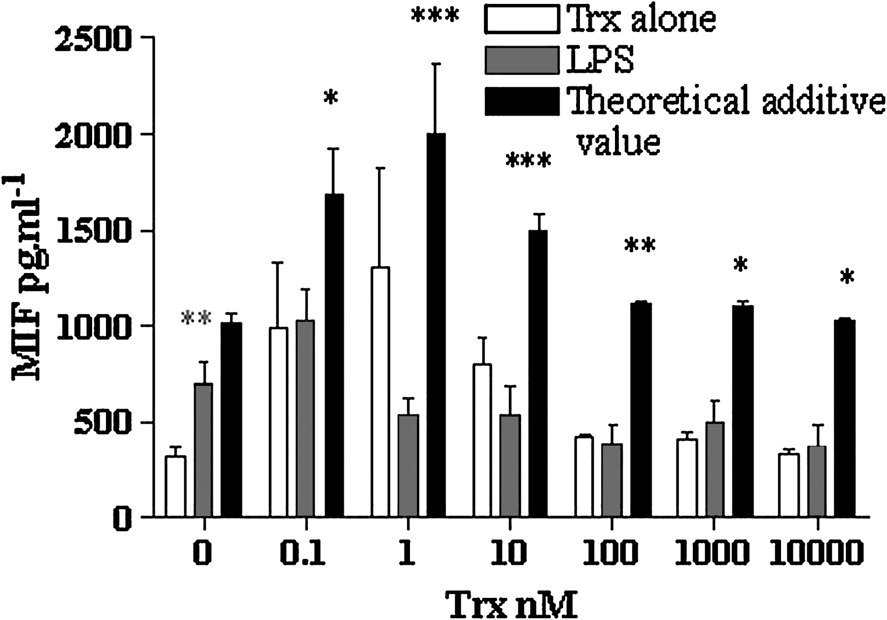
Results Following incubation with Trx there was no significant change in MIF release from monocytes. By contrast, LPS and LTA significantly (p<0.01) induced MIF from base line. When monocytes were treated with LPS (Abstract S51 Figure 1) or LTA following pre-incubation with Trx, MIF release was significantly less than the theoretical additive effects of the two treatments alone. By contrast, although Trx significantly induced IL-8 and IL-10, Trx did not modulate LPS or LTA induced cytokine release.
Effect of Trx of on release of MIF from human monocytes following stimulation with LPS. Human monocytes (1×10⁶ ml−1) we re pre-incubated with Trx (0.1–10 000 mM) for 24 h followed by stimulation with medium alone (white bars) or LPS 1 μg/ml(red bars) for 24 h MIF concentrations in cell supematants were determined by sandwich ELISA. The black bars represent the theoretical additive values of Trx and LPS. Data represent mean ± SEM from 5 experiments (LPS) and 3 experiments nts (no treatment). *p<0.05, **p(0.01, ***p<0.001 when conditions compared (two way ANOVA with Bonferroni's post test). Red asterisks represent significant increase following LPS alone. Black asterisks represent significant difference between LPS and Trx with the theoretical additive value. MIF, macropage migration inhubitory factor; LPS, lipopolysac charide; LTA, lipoteichoic acid; Trx, Thioredoxin.
Conclusion Trx reduced MIF release following stimulation with LPS and LTA. Extracellular Trx exerts an anti-inflammatory effect in this model. The Trx/MIF axis should be explored as a potential route for therapeutic intervention in patients with sepsis.