Article Text

Download PDFPDF

Pulmonary scedosporium infection as a complication of infliximab therapy for ankylosing spondylitis
Free
  1. J C Ngai1,
  2. R Lam2,
  3. F W Ko1,
  4. K-W To1,
  5. D S Hui1
  1. 1
    Department of Medicine and Therapeutics, The Chinese University of Hong Kong, Prince of Wales Hospital, Shatin, New Territories, Hong Kong
  2. 2
    Department of Microbiology, The Chinese University of Hong Kong, Prince of Wales Hospital, Shatin, New Territories, Hong Kong
  1. Dr D S Hui, Department of Medicine and Therapeutics, The Chinese University of Hong Kong, Prince of Wales Hospital, 30-32 Ngan Shing St, Shatin, New Territories, Hong Kong; dschui{at}cuhk.edu.hk

Statistics from Altmetric.com

Request Permissions

If you wish to reuse any or all of this article please use the link below which will take you to the Copyright Clearance Center’s RightsLink service. You will be able to get a quick price and instant permission to reuse the content in many different ways.

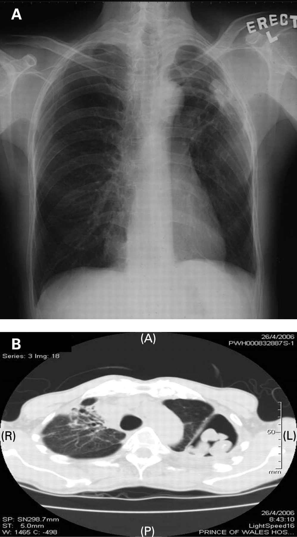
A 47-year-old man with a negative Mantoux test received infliximab 800 mg fortnightly in August 2005 for treatment of ankylosing spondylitis. He developed haemoptysis after receiving the third dose in September 2005 and therefore infliximab was stopped. CT thorax in April 2006 showed evidence of mycetoma at the left upper lobe (fig 1A, B). In view of persistent haemoptysis and radiological deterioration, voriconazole 200 mg daily was started and left upper lobe lobectomy was performed. Histopathology showed caseous granulomatous inflammation, destroyed bronchial wall with inflammatory cell infiltration and fungal mycelium was seen. Culture of the resected tissue grew Scedosporium apiospermum (fig 2A, B). Haemoptysis subsided after surgery.

Figure 1 (A) Chest x ray showed a small and non-specific opacity at the left upper lobe (LUL). His chest x ray prior to commencement of infliximab was unremarkable. (B) Chest CT revealed a cavity at the posterolateral aspect of LUL with several opacities within, suggestive of mycetoma. Inflammatory changes were also noted at the right upper lobe.
Figure 2 (A) Three fungal balls removed from the left upper lobe cavity. (B) Scedosporium apiospermum is the species of hyaline molds commonly encountered in clinical practice. Microscopically, hyaline mold is characterised by the formation of conidia singly from the tip of conidiophores. The conidiophores of S apiospermum are straight and narrow with no swelling. Some conidia aggregated into bundles in the background.

Learning points:

  • Scedosporium infection may present in a wide range of pulmonary manifestations from simple colonisation to mycetoma formation and invasive disease, simulating aspergillosis.1

  • Antitumour necrosis factor agents should be used with caution as these may lead to opportunistic infections as a result of suppression of T cell mediated immunity.2 To the best of our knowledge, this is the first case report of pulmonary scedosporium infection as a complication of infliximab therapy for ankylosing spondylitis.

REFERENCES

View Abstract

Footnotes

  • Competing interests: None.

  • Ethics approval: Ethics approval was obtained.

  • Patient consent: Obtained.

Linked Articles

  • Airwaves
    Wisia Wedzicha