Article Text

Statistics from Altmetric.com
CLINICAL PRESENTATION
A 41-year-old heterosexual man with a recently diagnosed HIV infection was admitted to our department complaining of a 5-month history of malaise, non-productive cough and a weight loss of 8 kg. He was a non-intravenous drug user.
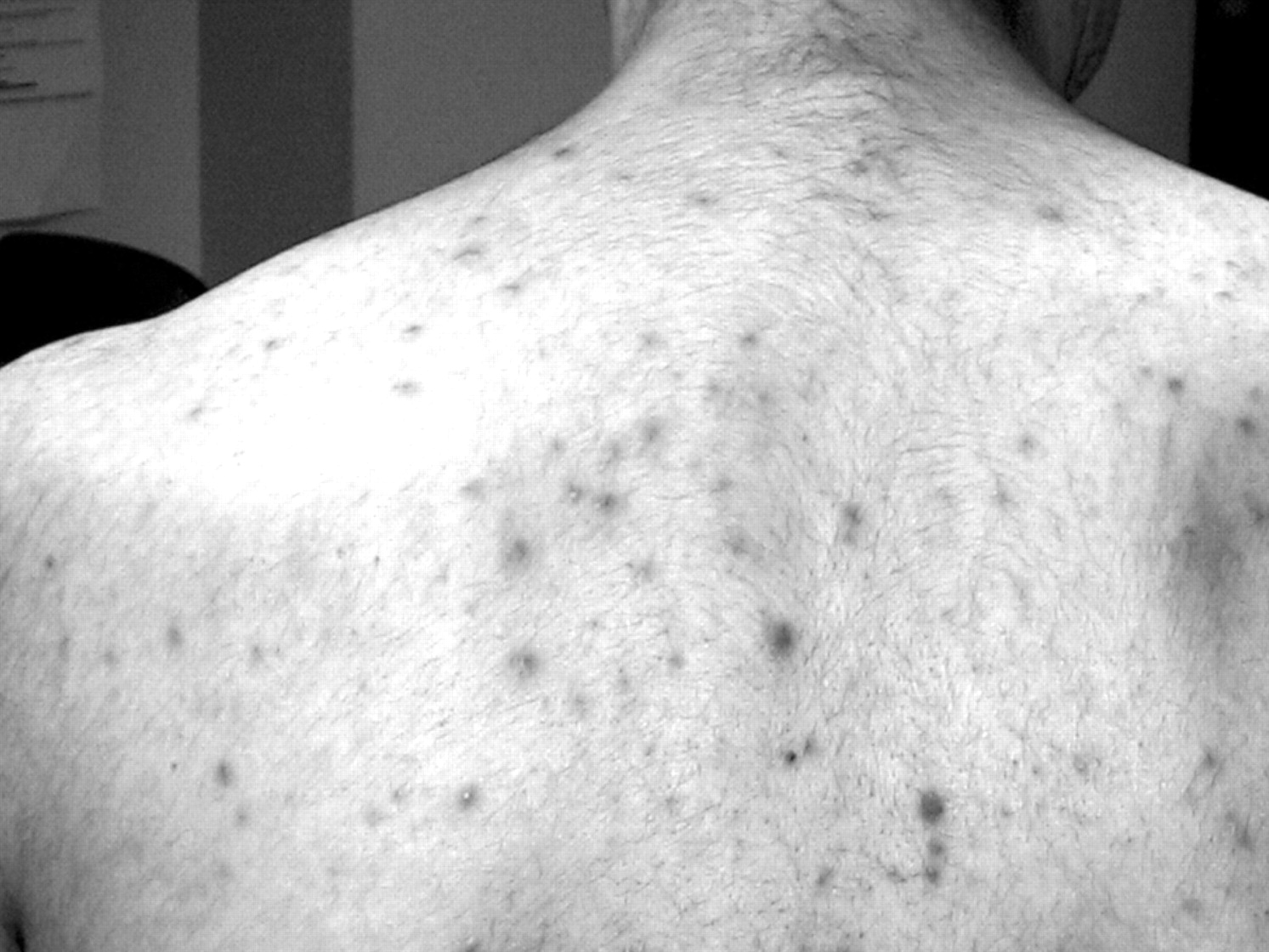
On physical examination he had a generalised maculopapular rash which was more prominent on the trunk and arms and included the palms and soles (fig 1). Physical examination of the heart, lungs and abdomen was normal. The CD4 count was 176 cells/μl and the plasma HIV-RNA was 4.3 log copies/ml. The results of blood screening laboratory tests were normal.
Chest radiography showed multiple bibasilar pulmonary nodules while a CT scan of the lungs showed a wide area of increased density with irregular margins located in the right inferior pulmonary lobe (fig 2). A tuberculin skin test was negative.

Cytological examinations of bronchoalveolar lavage (BAL) fluid did not show malignant cells. Gram, PAS, Ziehel-Neelsen and Warthin-Starry stains of specimens from lung brushing and transbronchial biopsy did not reveal infectious agents. Cultures of BAL fluid specimens for standard bacteria, mycobacteria and fungi were negative. Repeated haemocultures performed in the absence of concomitant antimicrobial therapy yielded negative results. A CT-guided transthoracic biopsy of the largest lung lesion showed an inflammatory infiltrate with numerous lymphocytes, granulocytes and histiocytes, without evidence of malignancy, fungal or mycobacterial infections.
QUESTIONS
What further analyses might be helpful? What is your diagnosis?
See page 173
This case was submitted by:
Footnotes
Competing interests: None.
Patient consent: Obtained.