Article Text

Statistics from Altmetric.com
A 56-year-old woman was admitted to our hospital with inspiratory dyspnoea, dry cough, vague right chest pain and right arm pain for 2 months. She denied tobacco use or asbestos exposure and had no significant medical or family history.
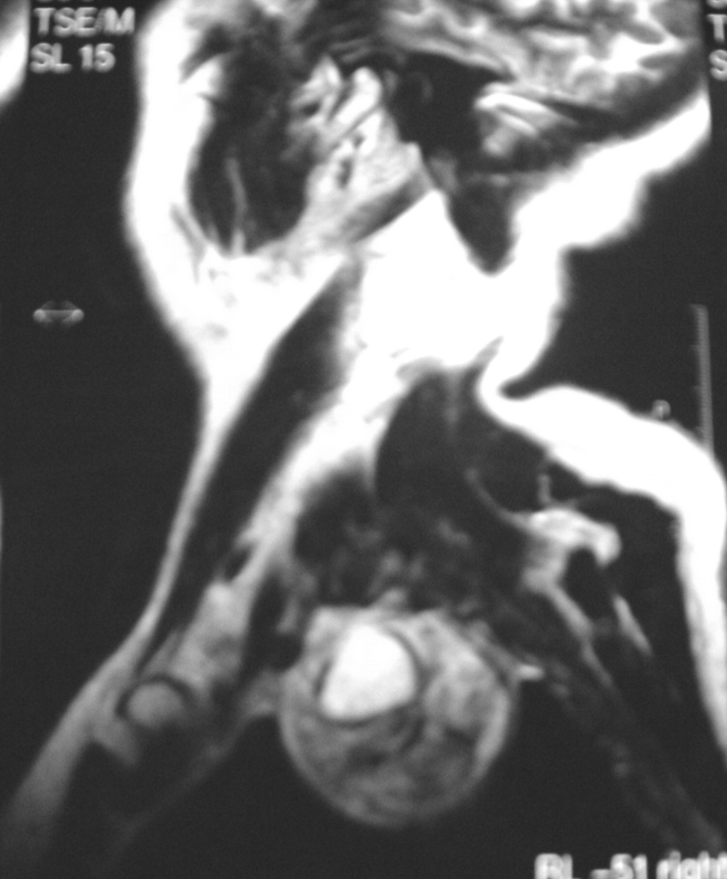
A chest radiograph highlighted extensive radiopacity involving the superior right lobe, the lateral view detailed the mass as being located in the posterior mediastinum. Chest CT scanning revealed a large well circumscribed heterogeneous tumour of 7×6×5 cm, with punctuate calcifications and focal low density areas, located in the posterior mediastinum in the right paravertebral gutter (fig 1). MRI demonstrated a well defined lobulated mass and inhomogeneous enhancement with non-enhanced linear and patchy areas after intravenous gadolinium injection. There were no expansions through spinal roots or other tissues (fig 2). The sample taken by CT guided fine needle aspiration of the mass was of equivocal value because it showed atypical features. The diagnosis of malignant tumour was assumed, and the patient underwent surgical exploration to determine the final histological diagnosis. The tumour, being of extrapleural origin, arising from intercostal nerve and having no intracanalicular extension, was isolated with a complete en bloc surgical resection. The histological diagnosis was “ancient schwannoma” (fig 3). The postoperative course was uneventful and the patient was discharged home on postoperative day 6.


The intercostal ancient schwannoma can mimic pulmonary neoplasm in chest radiographs and CT.1 MRI is the technique of choice in the assessment of peripheral nerve tumours.2 Ancient schwannoma is a rare variant of a neurilemoma and shows degenerative histological changes which may lead to a mistaken diagnosis of malignant neoplasm. The definitive diagnosis is possible only after histopathological examination.
Learning points
Ancient schwannoma is a rare variant of a neurilemoma.
The intercostal ancient schwannoma can mimic pulmonary neoplasm in chest radiographs and CT.
Ancient schwannoma shows degenerative histological changes which may lead to a mistaken diagnosis of malignant neoplasm.
Footnotes
Competing interests: None.
Patient consent: Obtained.
Linked Articles
- Airwaves